Common Ultrasound Applications
What is ultrasound best used for?
- Superficial structures: vessels, palpable masses, etc
- Solid organs: liver, kidneys, thyroid
- Pediatrics: spares children radiation, less ossification of cartilagenous structures allows better imaging (ie: spine, brain)
Applications
- Gallbladder and renal stones
- Echocardiography
- Solid organ mass evaluation
- Appendicitis, especially in young patients
- FAST exam in trauma
- Vascular diseases
- Obstetrical imaging
- Some chest and intracranial applications, but limited by air in the lungs and bony structures (calvarium)
- Guidance for interventional percutaneous, endocavitary, and intraoperative/laparoscopic procedures
- And many more …
Right Upper Quadrant - Cholelithiasis
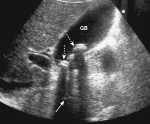
Here we see a gallbladder with some fluid and multiple shadowing calculi.
Retroperitoneal – renal calculi
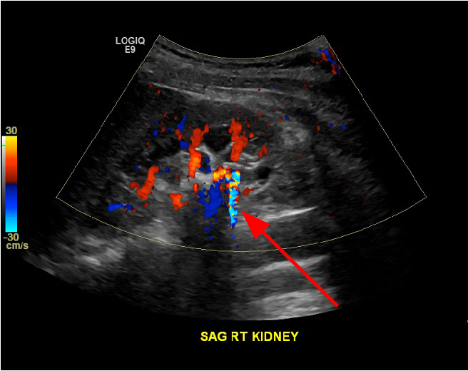
The image shows a retroperitoneal (kidneys, ureters and bladder) ultrasound focused on the right kidney with doppler signal, showing twinkle artifact over a kidney stone.
Appendicitis
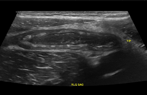
This is a right lower quadrant ultrasound of a child, which allows us to spare the child radiation. A dilated, thick walled, inflammed appendix is seen here, oriented transversely.
FAST Exam- Left Upper Quadrant
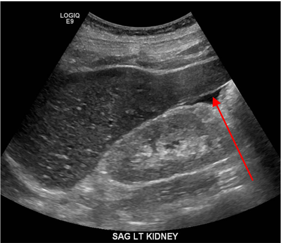
The FAST Exam is a bedside assessment tool used to assess for free fluid, such as blood, in the abdomen after a trauma. Here we see the left upper quadrant, with a small amount of free fluid (arrow) near the kidney.
Vascular imaging- DVT
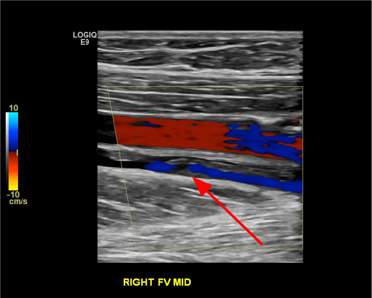
The image here shows vascular ultrasound, showing both the right femoral vein and artery. The vein has echogenic material within it (arrow), with no color doppler signal in the area of the material, which is a clot, or a deep vein thrombosis
Obstetrical
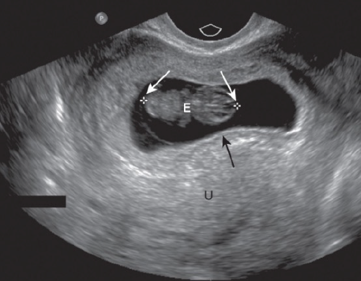
Fetus- intrauterine pregnancy. The white arrows point to the measurement locations for "crown rump length" used to measure fetal size. The black arrow points to the edge of the gestational sac. Radiology departments generally only read 1st trimester ultrasound, and obstetrics will perform and read 2nd and 3rd trimester ultrasound
Neonatal brain ultrasound
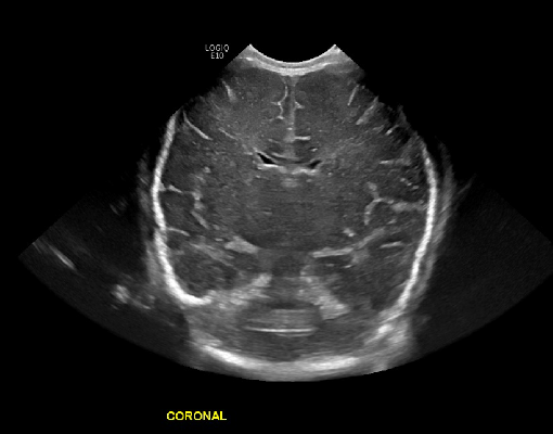
The image here shows a head ultrasound in a neonate. The fontanelles are a perfect window to ultrasound the developing brain. We have a coronally oriented image through the cerebral hemispheres and lateral ventricles.
Breast biopsy
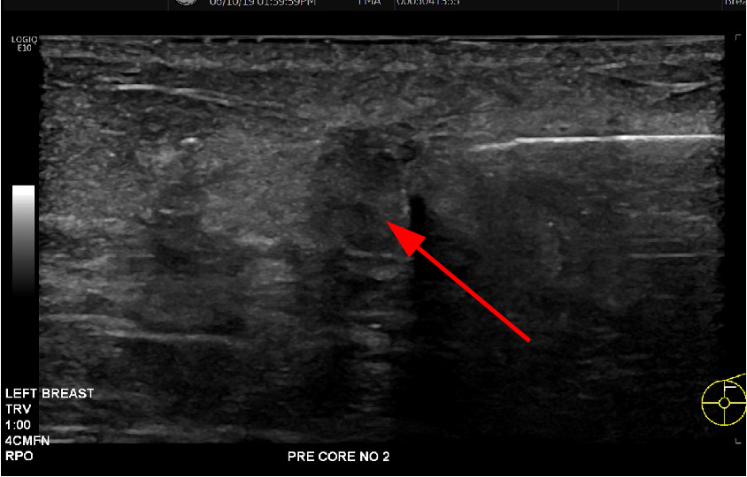
The image here shows ultrasound guidance for a biopsy of a hypoechoic breast mass. The needle is the hyperechoic structure, shown here just prior to entering the mass (arrow)