Ultrasound Equipment
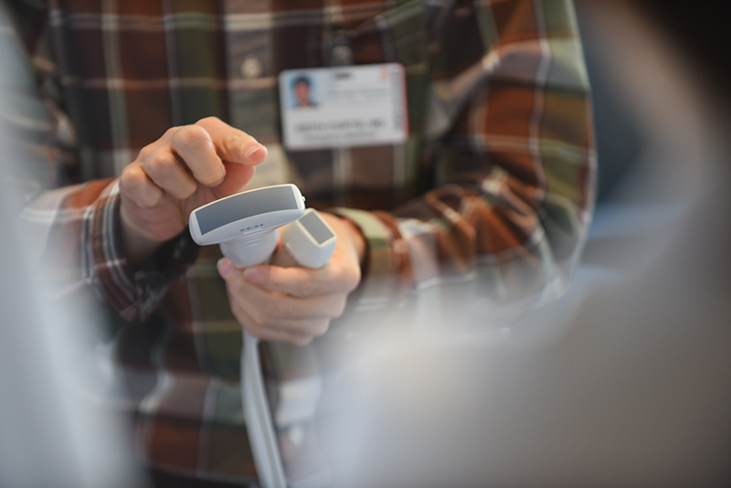
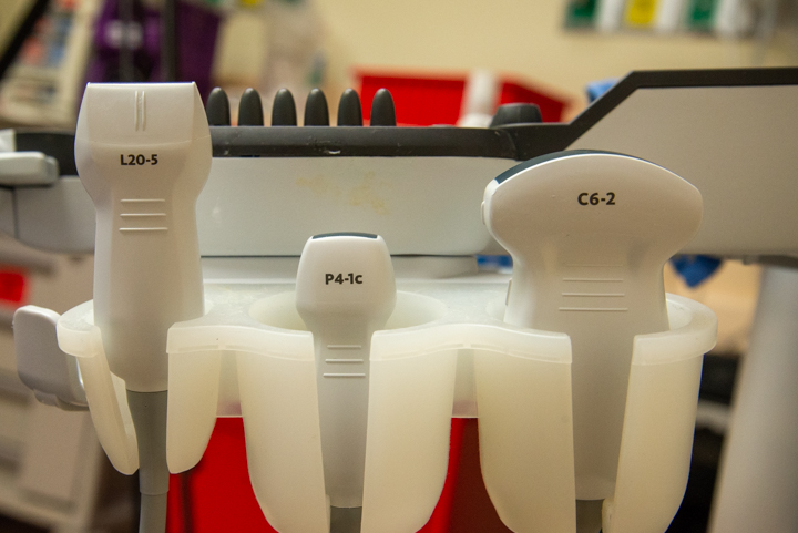
Types of Probes

The various types of transcutaneous probes. All of these have piezoelectric crystals.
Probes have different frequency ranges that they can be used with. High frequencies do not penetrate tissues deeply, so they are good at looking at superficial structures. An example of this type of probe is the linear array probe, frequently used to look at vasculature. Curved array and phased array probes have lower frequency ranges, which can look at deeper structures, but have lower resolution images.
Equipment Settings
M-Mode
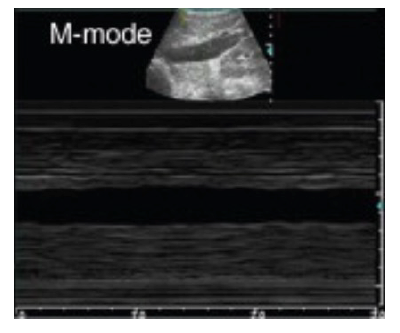
Different settings on the machine can be used to make the images appear differently. M-mode, for example, displays a line of motion over time, showing how objects move relative to the receiver. This is useful for examining this such as a fetal heart beat or lung sliding.
Depth and Gain
Gain adjusts the amount of sound being picked up by the receiver, and does not impact sound waves going out. Many machines will have separate time gain compensation knobs for near and far field, which allows you to change how bright different depths of objects appear.
Focus changes the depth of the focus of the ultrasound waves, allowing imaging of objects at different depths.
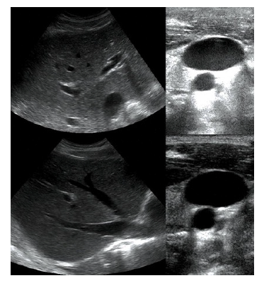
Doppler
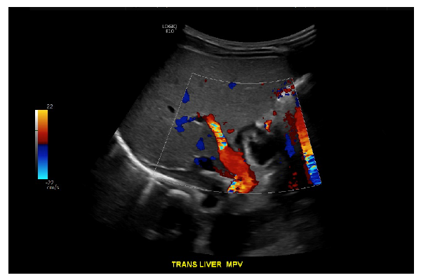
Doppler uses information about how quickly sound waves are returning to the probe to determine if there is movement away from or towards the probe. This is useful with fluids, especially in vascular imaging.
Its important to recognize that the blue/red color scheme does not reflect arterial or venous blood, simply direction of flow. Here we have a doppler image of the liver, with red flow in the main portal vein indicating flow towards the transducer and therefore towards the liver.
Using an ultrasound machine (video)
![]()