Common Ultrasound Artifacts
Twinkle Artifact
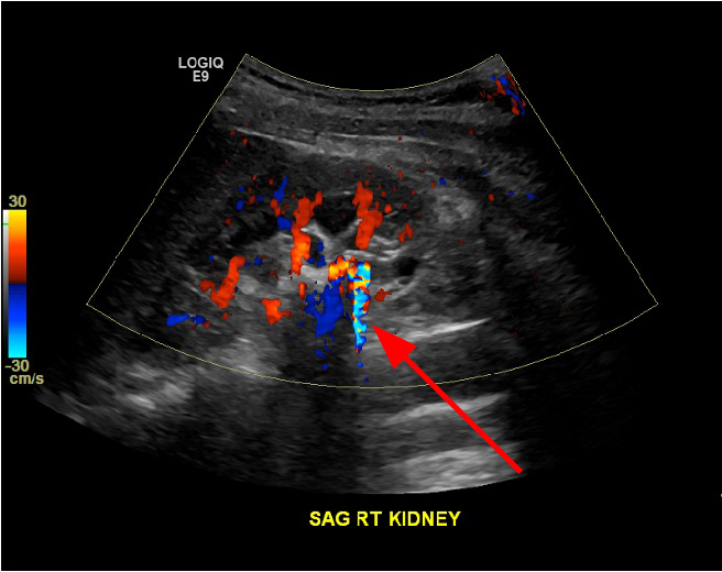
- Occurs with doppler on calculi and other rough surfaces
- Also called scintillation
- Physics behind this is poorly understood
Here we see a gallbladder with a calculus in it. When doppler signal is put on this area, mixed shimmering colors are seen shining down from the calculus, called "twinkle artifact." The physics behind this is poorly understood, but most likely is related to sound waves reverberating off of a rough surface.
Reverberation Artifact
- When sound hits two parallel high reflective surfaces, the signal appears to "reverberate" deep to the surfaces
- Occurs with metal or glass foreign bodies
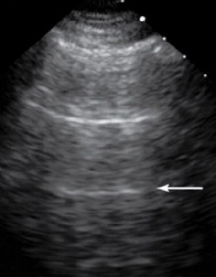
The image here shows lung ultrasound. Its important to note that we do not see normal lung well with ultrasound, as it is filled with air and air is a poor acoustic medium, as discussed previously. Here, we see reverberation of sound reflecting off of the visceral and parietal pleura, which are are highly reflective. The arrows point to repetition of this signal.
Mirror Image Artifact
- When sound hits a single highly reflective surface, the sound returns to the transducer more quickly than expected
- This causes "mirroring" of the signal on the opposite side of the reflective surface
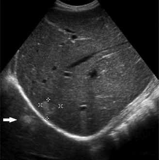
The image here shows a liver lesion which is mirrored on the opposite side of the diaphragm, a highly reflective surface.
Posterior Acoustic Shadowing
- When sound hits a single highly dense surface, the sound is all reflected, with no signal passing beyond the surface
- This occurs with bone and calculi
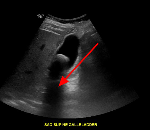
Here we have an image of a large calculus in the gallbladder, which causes shadowing posterior to the most anterior surface of the stone
Posterior Acoustic Enhancement
- Sound travels very quickly through certain structures such as fluid, and when this occurs, the area behind it appears "bright" or "enhancing"
- This occurs with fluid filled structures such as the gallbladder
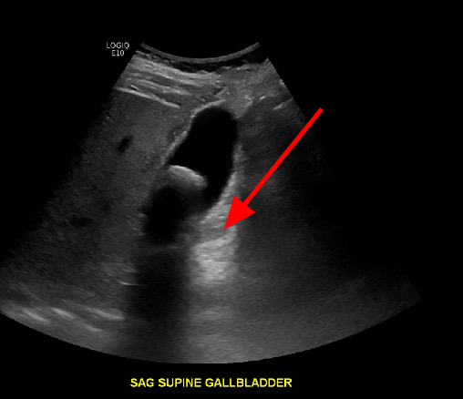
The reason this occurs is again that the transducer receives sound waves more quickly than expected and this causes the appearance of brighter or increased echogenicity. Here we have the same image of the gallbladder, with enhancement posterior to the fluid filled gallbladder.