Ultrasound
Introduction to Ultrasound
Lesson Objectives
- Describe the basic physics of ultrasound
- Recognize the different ultrasound probes and settings (i.e. doppler, M-mode, depth, focus)
- Compare and contrast echogenic vs. hypoechoic structures
- Identify common ultrasound artifacts
- Recognize the strengths and weaknesses of ultrasound imaging
How are Ultrasound images obtained
 Ultrasound is so named because it uses waves above the range of human hearing (>20,000 Hz), "ultrasonic." Ultrasound uses special probes with piezoelectric quartz crystals. This means that the crystals are able to change electrical energy into mechanical energy (sound waves), send those waves into the body, and then receive waves coming back that have bounced off objects in the body, and convert them back into electrical signals which can be displayed on a screen. Ultrasound probes use the time that it takes for waves to return and the frequency of the waves to returning to create an image with different areas of light and dark.
- Transmit sound waves into the body
- Sound reflected by various tissues
- Reflected sound is captured and converted into an image
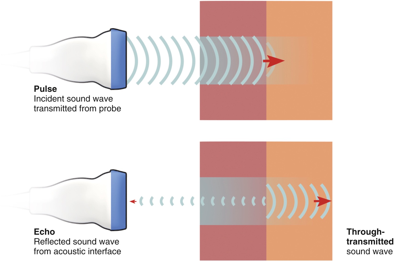
Some sound is reflected (echoed) when the ultrasound pulse encounters an interface between tissues with different values of acoustic impedence. The remaining unreflected sound energy continues on (through-transmission).
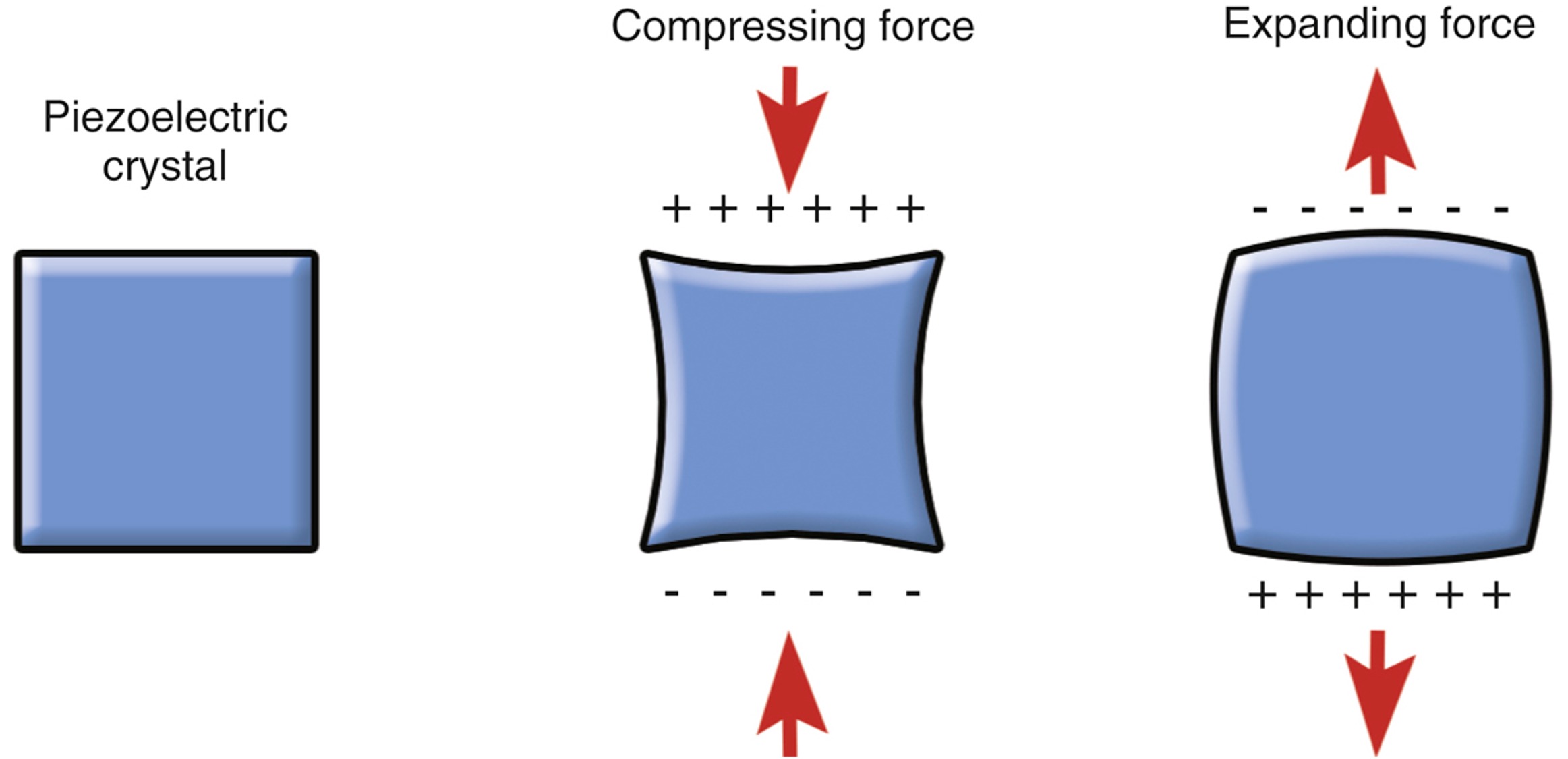
Mechanical deformation of piezoelectric crystals results in a net charge across the crystal.
How is this different from Radiographs?
- No tissue damage in the diagnostic range of frequencies
- Real-time
- Portable
- Ultrasound has no ionizing radiation that may harm the patient
The following video demonstrates the basic functionality of an ultrasound machine.
Ultrasound Equipment
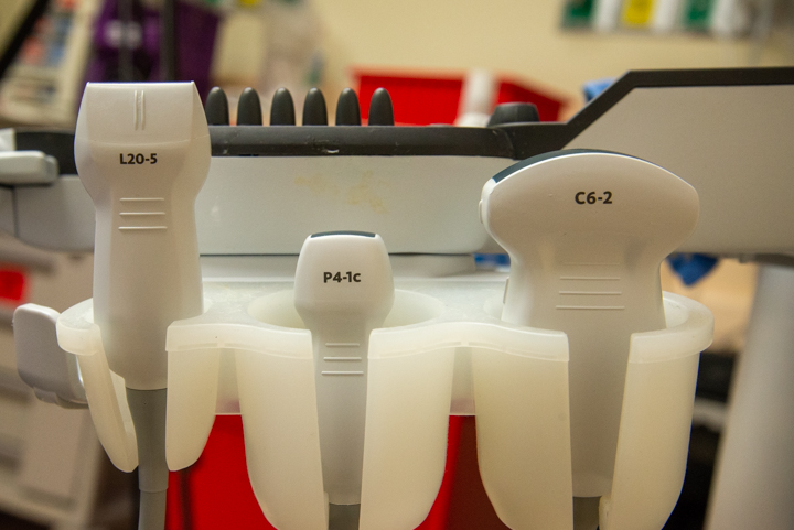
Types of Probes

The various types of transcutaneous probes. All of these have piezoelectric crystals.
Probes have different frequency ranges that they can be used with. High frequencies do not penetrate tissues deeply, so they are good at looking at superficial structures. An example of this type of probe is the linear array probe, frequently used to look at vasculature. Curved array and phased array probes have lower frequency ranges, which can look at deeper structures, but have lower resolution images.
Equipment Settings
M-Mode
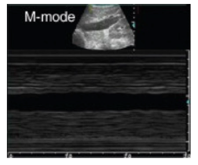
Different settings on the machine can be used to make the images appear differently. M-mode, for example, displays a line of motion over time, showing how objects move relative to the receiver. This is useful for examining this such as a fetal heart beat or lung sliding.
Depth and Gain
Gain adjusts the amount of sound being picked up by the receiver, and does not impact sound waves going out. Many machines will have separate time gain compensation knobs for near and far field, which allows you to change how bright different depths of objects appear.
Focus changes the depth of the focus of the ultrasound waves, allowing imaging of objects at different depths.
Doppler
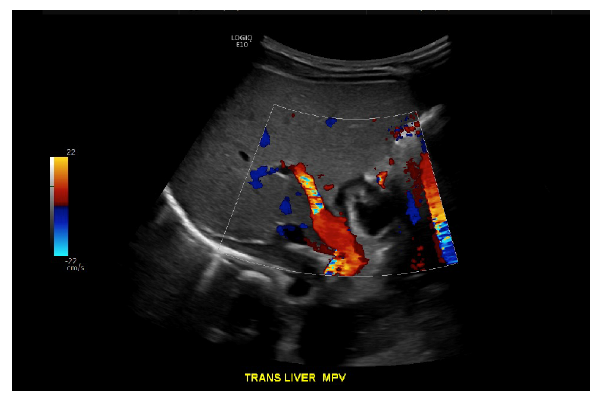
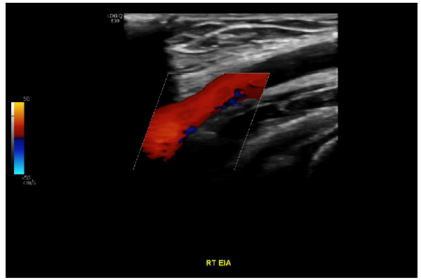
Doppler uses information about how quickly sound waves are returning to the probe to determine if there is movement away from or towards the probe. This is useful with fluids, especially in vascular imaging.
Its important to recognize that the blue/red color scheme does not reflect arterial or venous blood, simply direction of flow. Here we have a doppler image of the liver, with red flow in the main portal vein indicating flow towards the transducer and therefore towards the liver.
Using an ultrasound machine (video)
Quiz - Test your knowledge
What makes things bright or dark on ultrasound?
- Bright on ultrasound = hyperechoic / echogenic
- Dark on ultrasound = hypoechoic
- Black on ultrasound = anechoic
The words that we use to describe things that are bright or dark on ultrasound correspond with the physics. Higher intensity reflected sound waves are "hyperechoic" or "bright," and vice versa. Things that are anechoic have a complete absence of returning sound waves.
Acoustic impedance
This is probably a flashback to high school physics, but we'll have to review acoustic impedance to further understand echogenicity. Acoustic impedance in the body is a product of the density of the tissue and the velocity of sound in the medium.
Based on these principles, sound travels most quickly and well through solid and especially fluid, and not well through air (think of knocking on a door or whales comminicating from miles away underwater). When sound waves encounter an interface where there is a large difference in acoustic impedance, they are reflected, which causes a failure in sound waves to travel deeper into the tissue.
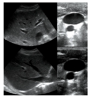
Anechoic - fluid (urine) in bladder.
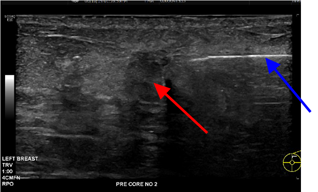
Hypoechoic - breast mass (red arrow), hyperechoic - needle (blue arrow.)
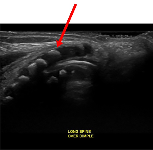
Hyperechoic - bone (neonatal spine.)
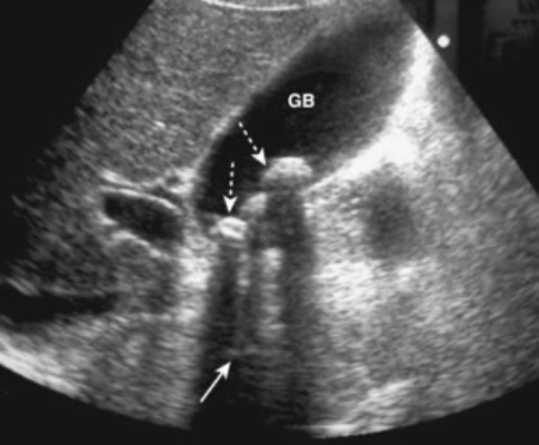
Common Ultrasound Artifacts
Twinkle Artifact
- Occurs with doppler on calculi and other rough surfaces
- Also called scintillation
- Physics behind this is poorly understood
Here we see a gallbladder with a calculus in it. When doppler signal is put on this area, mixed shimmering colors are seen shining down from the calculus, called "twinkle artifact." The physics behind this is poorly understood, but most likely is related to sound waves reverberating off of a rough surface.
Reverberation Artifact
- When sound hits two parallel high reflective surfaces, the signal appears to "reverberate" deep to the surfaces
- Occurs with metal or glass foreign bodies
The image here shows lung ultrasound. Its important to note that we do not see normal lung well with ultrasound, as it is filled with air and air is a poor acoustic medium, as discussed previously. Here, we see reverberation of sound reflecting off of the visceral and parietal pleura, which are are highly reflective. The arrows point to repetition of this signal.
Mirror Image Artifact
- When sound hits a single highly reflective surface, the sound returns to the transducer more quickly than expected
- This causes "mirroring" of the signal on the opposite side of the reflective surface
The image here shows a liver lesion which is mirrored on the opposite side of the diaphragm, a highly reflective surface.
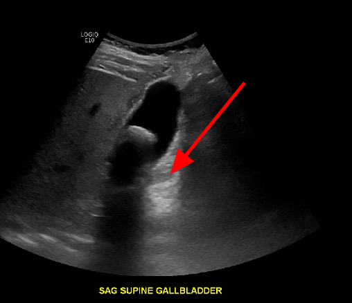
Posterior Acoustic Shadowing
- When sound hits a single highly dense surface, the sound is all reflected, with no signal passing beyond the surface
- This occurs with bone and calculi
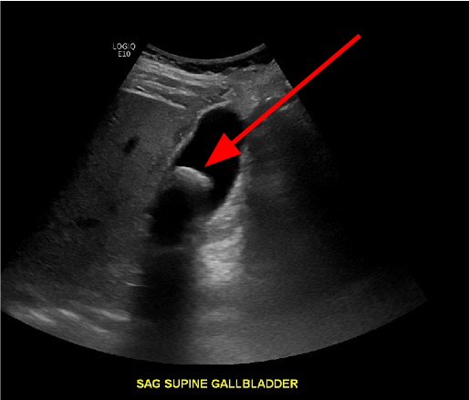
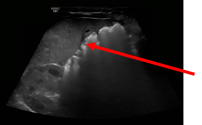
Here we have an image of a large calculus in the gallbladder, which causes shadowing posterior to the most anterior surface of the stone
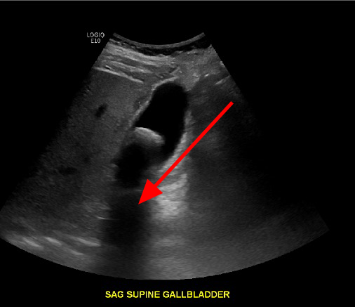
Posterior Acoustic Enhancement
- Sound travels very quickly through certain structures such as fluid, and when this occurs, the area behind it appears "bright" or "enhancing"
- This occurs with fluid filled structures such as the gallbladder
The reason this occurs is again that the transducer receives sound waves more quickly than expected and this causes the appearance of brighter or increased echogenicity. Here we have the same image of the gallbladder, with enhancement posterior to the fluid filled gallbladder.
Common Ultrasound Applications
What is ultrasound best used for?
- Superficial structures: vessels, palpable masses, etc
- Solid organs: liver, kidneys, thyroid
- Pediatrics: spares children radiation, less ossification of cartilagenous structures allows better imaging (ie: spine, brain)
Applications
- Gallbladder and renal stones
- Echocardiography
- Solid organ mass evaluation
- Appendicitis, especially in young patients
- FAST exam in trauma
- Vascular diseases
- Obstetrical imaging
- Some chest and intracranial applications, but limited by air in the lungs and bony structures (calvarium)
- Guidance for interventional percutaneous, endocavitary, and intraoperative/laparoscopic procedures
- And many more …
Right Upper Quadrant - Cholelithiasis
Here we see a gallbladder with some fluid and multiple shadowing calculi.
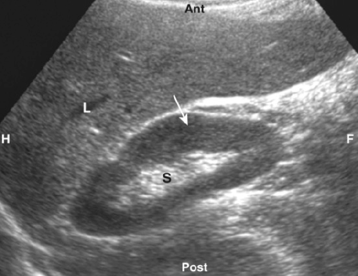
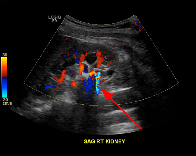
Retroperitoneal – renal calculi
The image shows a retroperitoneal (kidneys, ureters and bladder) ultrasound focused on the right kidney with doppler signal, showing twinkle artifact over a kidney stone.
Appendicitis
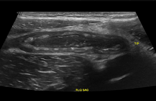
This is a right lower quadrant ultrasound of a child, which allows us to spare the child radiation. A dilated, thick walled, inflammed appendix is seen here, oriented transversely.
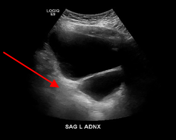
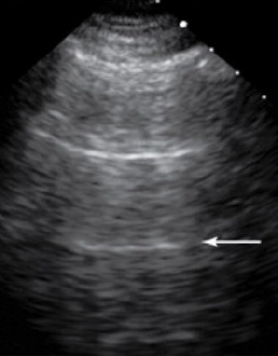
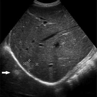
FAST Exam- Left Upper Quadrant
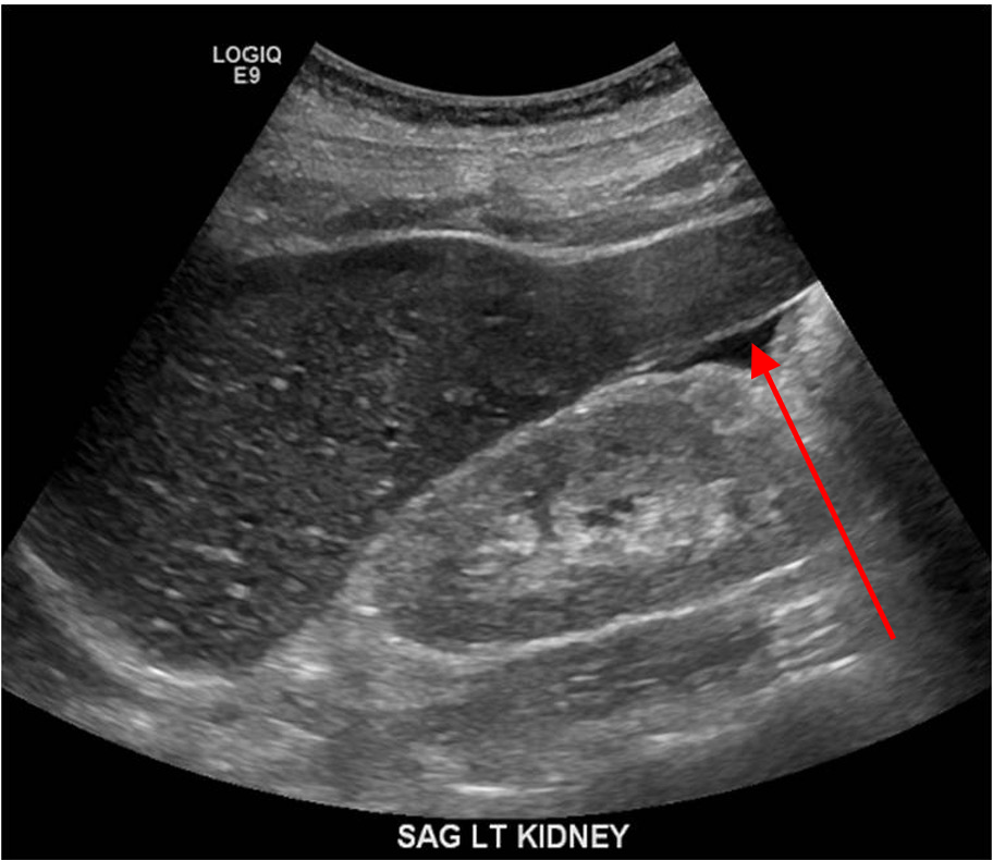
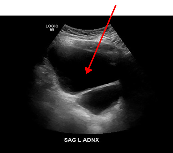
The FAST Exam is a bedside assessment tool used to assess for free fluid, such as blood, in the abdomen after a trauma. Here we see the left upper quadrant, with a small amount of free fluid (arrow) near the kidney.
Vascular imaging- DVT
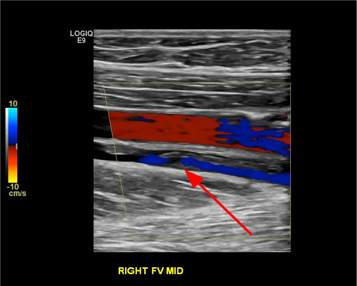
The image here shows vascular ultrasound, showing both the right femoral vein and artery. The vein has echogenic material within it (arrow), with no color doppler signal in the area of the material, which is a clot, or a deep vein thrombosis
Obstetrical
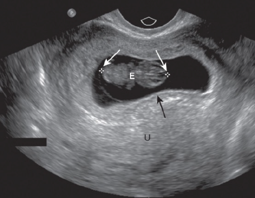
Fetus- intrauterine pregnancy. The white arrows point to the measurement locations for "crown rump length" used to measure fetal size. The black arrow points to the edge of the gestational sac. Radiology departments generally only read 1st trimester ultrasound, and obstetrics will perform and read 2nd and 3rd trimester ultrasound
Neonatal brain ultrasound
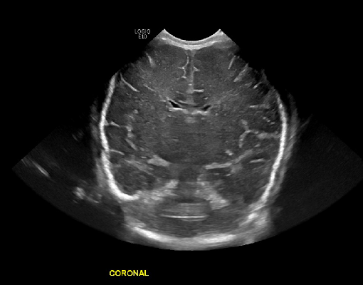
The image here shows a head ultrasound in a neonate. The fontanelles are a perfect window to ultrasound the developing brain. We have a coronally oriented image through the cerebral hemispheres and lateral ventricles.
Breast biopsy
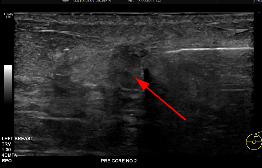
The image here shows ultrasound guidance for a biopsy of a hypoechoic breast mass. The needle is the hyperechoic structure, shown here just prior to entering the mass (arrow)
Quiz - Test your knowledge