Protein Structure


Protein Structure amino acids primary structure secondary structure tertiary structure quaternary structure
Protein Function Structure, SHAPE, and Function of Tools Structure, SHAPE, and Function of Proteins Arabinose Transporter Hexokinase


Amino Acids  -  the subunits of the polypeptide chain.

Every amino acid has the same structure because an amino group is always linked to a carboxyl group in all amino acids
However, amino acids have different structures because the R group (or side chain) is different for each kind of amino acid. 

This image shows the 20 amino acids which make up proteins.

  • The part which is the same in all amino acids is shown in red.
  • The part which is different in all amino acids is shown in blue.
  • Proteins (or polypeptides) are chains of amino acids linked together by peptide bonds through their carboxyl and amino groups.  The side chains stick out to the side.
    • An average protein is on the order of 150 amino acids long, although some are shorter (Insulin), and many are longer.
    • Since there are over 100 amino acids in the polypeptide chain, and at each position there can be any one of 20 different amino acids, there are millions of different SEQUENCES possible.


    Primary Structure   -   the sequence of amino acids in the peptide chain.
    •  All proteins are composed of a chain of amino acids. 
    • There are millions of different SEQUENCES possible.  For example:
      1. ala-trp-cys-ser-his-trp-trp-gly-glu-ileu   is one sequence.
      2. ser-his-trp-trp-ala-trp-cys-gly-glu-ileu   is another sequence.
      3. trp-gly-glu-ileu-ala-trp-cys-ser-his-trp  is a third sequence.
    • Every protein has a characteristic sequence of amino acids .......  this is the primary structure of that particular protein.  For example:
      1. the first sequence might be  the first 10 amino acids found in all molecules of phenylalanine hydroxylase.
      2. the second sequence might be  the first 10 amino acids found in all molecules of tyrosine hydroxylase.
      3. the third sequence might be  the first 10 amino acids found in all molecules of Homogentisic Acid Oxidase.


    The primary structure determines what secondary structures will be formed!!


    Secondary Structures  occur when the primary structure (the amino acid chain) folds into various secondary conformations such as alpha helix, beta pleated sheet oe random coil. 

    The primary structure determines what secondary structures will be formed!!

     
    a helix is formed when the amino acid chain spirals upward in the form of a right-handed helix.  In this image, each amino acid is circled.  It can be seen that the amino acids are chained together - but then the chain coils up!

    A stereo picture gives a better idea of the 3-D coiling in a helix.  The amino acid chain twists into this shape because weak charge attractions (hydrogen bonds) form between different amino acids. In this picture, the dotted lines are hydrogen bonds
     

    HINT:  To see this is stereo,  put your nose up to the screen so you can't focus on either image.  Move slowly back .... DO NOT allow your eyes to focus!  You will quickly notice 3 images.  Concentrate on the middle one as you continue moving back.  At a certain point, depending on your own eyesight, the central one will come into focus, and appear to be three-dimensional!
    b pleated sheet is formed when segments of the amino acid chain run opposite to each other in an antiparallel orientation. A stereo picture gives a better idea of the 3-D shape in b pleated sheet.
    Random coil  is formed when a stretch of the amino acid chain does not form into either an a helix or a b pleated sheet.  In this image of whale myoglobin, you can see:
    1. Primary structure:  each amino acid is numbered from 1 - 153.
    2. Secondary structure:  both the a helix and random coil are easily seen.


    The secondary structure determines what tertiary structures will be formed!!


    Tertiary Structures are formed as follows:
    • The primary structure of a polypeptide is a chain of amino acids in a very specific sequence which can fold up to form any of the 3 kinds of secondary structure.
    • The different elements of secondary structure can then "pack" together to form a 3-D SHAPE This is called the tertiary structure of the protein. 
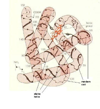

    Whale Myoglobin.  An example which shows how secondary structures form tertiary structure is whale myoglobin. This protein is similar to human b globin and binds oxygen. It is found in the muscles of all vertebrates and serves as a storage mechanism for oxygen between the time it is delivered to the muscle cell by the blood and the time it is used in oxidative phosphorylation.  The tissues of whales are loaded with this protein, and it is one reason that they can stay beneath the surface for nearly an hour. The rusty red structure is the porphyrin ring which traps an iron atom.  Since iron is easily oxidized (as in iron oxide or rust), it complexes with the oxygen absorbed from the blood, and "stores" it until it is used by the muscles.
     

    3 views of whale myoglobin tertiary structure:

    • In the drawing of whale myoblobin above, it can be seen that a SHAPE is formed when the different regions of a helix fold up on each other. This SHAPE is a third level of structure - tertiary structure. 
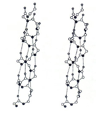
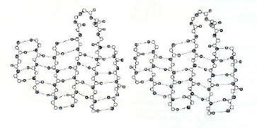
    • A different way of depicting the whale myoglobin is shown in the stereo cartoon image of whale myoglobin. This shows how the tertiary structure is formed from 2 different kinds of secondary structure; random coil is the gray strand and a helix is shown by the magenta coils.
      • NOTE: View this as a stereo image and note that the folding creates a 3D structure!
    • Another way of depicting whale myoglobin is shown in the stereo space filling image of whale myoglobin (this is the same molecule, in the same orientation, with the same color coding).  Here, it can be seen how the tertiary structure produces a SHAPE.
    The Tertiary Structure determines the SHAPE of the protein!


    Quaternary Structure.   A single protein may be made up of more than one amino acid chain:
    • dimers (2 amino acid chains - or peptide chains)
    • trimers (3 peptide chains)
    • tetramers (4 peptide chains)
    • pentamers (5 peptide chains)
    • hexamers (6 peptide chains)
    • etcetera !!
    Hemoglobin is a tetramer.  In this space-filling model of hemoglobin each amino acid chain is shown in a different color ( light blue in back; green on the right; yellow green to the left; blue in the foreground-lower left.  The small rusty red molecules inserted into the individual polypeptides and bound there (the other 2 in the back and can't be seen), are the porphyrin rings which bind the oxygen.


    In summary, what we have said is this:


    What is so important about SHAPE?


    RETURN to the  Protein Index Page

    Go to the top of the page.