Common Clinical Applications
The following are examples of common clinical applications of nuclear medicine.
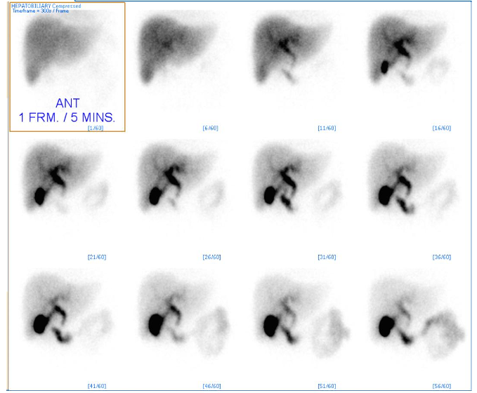
HIDA Scan
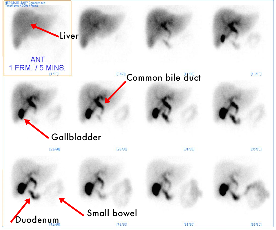
- HIDA (hepatobiliary iminodiacetic acid) scan- Similar to Bone scans, 2D planar images are used to visualize the amount of radiotracer uptake in the biliary tree, specifically the gallbladder.
- Tc-99m Mebrofenin, is taken up by the liver, then excreted into the biliary system where it collects in the gallbladder.
- Over time, it will be excreted via the common bile duct into the duodenum.
Why is this a useful test?
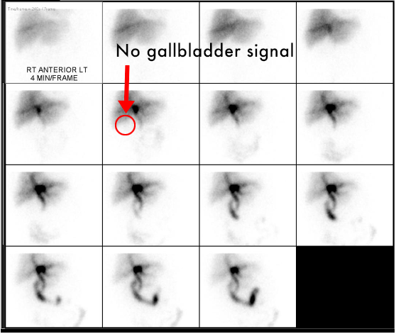
- Acute Cholecystitis: A LACK of radiotracer uptake within the gallbladder.
- Chronic cholecystitis: A gallbladder that fills with radiotracer, but does not empty appropriately after being given an emptying agent.
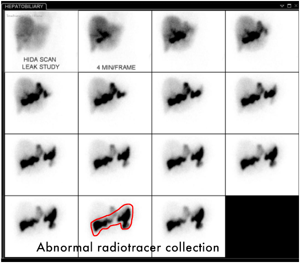
- Bile Leak: Radiotracer OUTSIDE of the biliary tree shortly following surgical removal of the gallbladder.
Acute Cholecystitis
- A normal HIDA scan will demonstrate radiotracer filling the gallbladder, as demonstrated on the previous images.
- In these abnormal images, the radiotracer is visible within the liver, the biliary tree, and the duodenum. However, there is no radiotracer uptake in the gallbladder.
- This is acute cholecystitis! BUT just to be sure (and to avoid unnecessary surgery if it isn't):
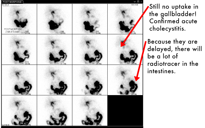
- Morphine administration will cause the Sphincter of Oddi to contract (where the common bile duct drains into the duodenum). The increased pressure in the CBD increases the likelihood of radiotracer entering the gallbladder.
Bile Leak
Shortly after undergoing cholecystectomy, the patient has continued right upper quadrant pain, fevers… The surgeon is concerned about a leak…
- CT scan may or may not give you the answer, because there can be normal postoperative fluid collections that may hide a small bile leak.
- HIDA scan demonstrates radiotracer OUTSIDE of the biliary tree, and not following the course of the intestines.
SPECT-CT - Bone Scan
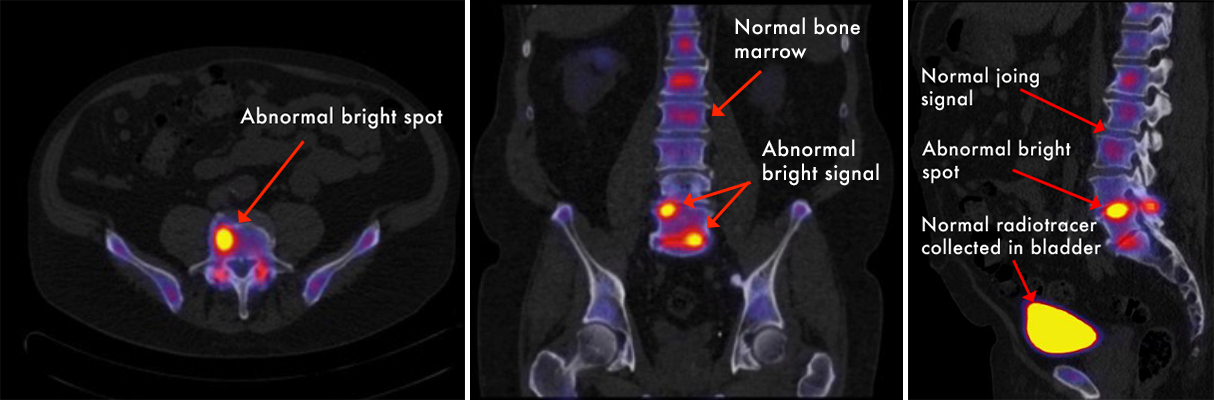
- SPECT = Single Photon Emission Computed Tomography
- Similar in principle to PET-CT, but utilizes different radiotracers to produce gamma rays instead of positrons.
- Primarily used to help target pain injections or guide therapy.
- Combination of CT and nuclear medicine images allow for improved resolution of images and provides anatomical localization.
- Abnormal signal will appear as bright "hot spots," indicating active inflammation and bony remodeling.
- Corresponding abnormalities on the CT images helps us give a more accurate diagnosis.
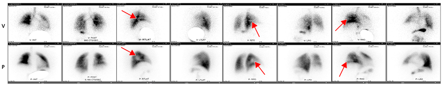
Ventilation/Perfusion Scan (VQ Scan)
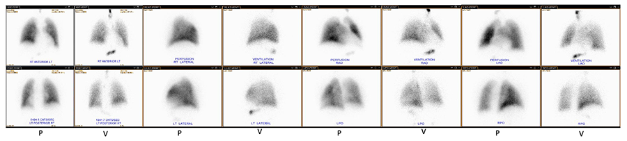
- Two sets of images are taken, with images taken from several different angles:
- The first scan uses inhaled radiotracer particles to demonstrate the airways and any ventilated lung tissue.
- The second scan uses a different injected radiotracer to demonstrate any perfused lung tissue.
- The two scans are compared to look for areas of mismatch. In a case with pulmonary embolism, there would be one or more regions of the lung that are ventilated, but not perfused because the PE is blocking the flow of blood to that area. Look for areas that don't match!
- CAUTION! Always check the labels on the images to see if it a perfusion (P) or ventilation (V) image- they may be organized in rows or columns.
Thyroid imaging: Iodine-123
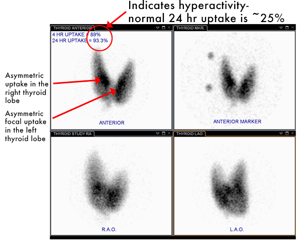
- The thyroid is very efficient at taking up iodine, relative to the rest of the body.
- Because of this, Iodine-123 is ideal for thyroid nuclear imaging.
- Planar images in the thyroid gland are taken at 4hr and 24hr. Normal uptake at 24 hours is typically 15-25% of the administered dose.
Why is this useful?
- I-123 scans can determine the degree of thyroid uptake and identify any focal or diffuse abnormal activity. For example...
- Graves Disease: thyroid stimulating immunoglobulin causes diffusely increased uptake.
- Toxic nodule: Asymmetric focal area of increased uptake, indicating a hyperactive nodule.
- Thyroid cancer: a thyroid nodule with a LACK of I-123 uptake is concerning for thyroid cancer.
Role of Radioisotopes in Treatment
I-131 is a radioisotope used for the treatment of thyroid cancer.
- Usually surgery is done prior to I-131 treatment with near total thyroidectomy (parathyroid glands usually preserved).
- Dose is usually given orally.
- Scan after surgery helps localize remnant thyroid tissue or metastases not identified prior to surgery. It also serves to ablate any residual thyroid tissue or any likely remnant metastatic cells.
- I-131 simultaneously images and treats and residual thyroid cancer. TTc