PET-CT is one of the most common studies performed in nuclear medicine with many applications.
Applications: Assist in staging, treatment response, and monitoring for recurrence for many cancers. Myocardial perfusion testing. Bone scans. And more.
FDG = fluoro-deoxy glucose, which is made using a radioactive isotope of fluorine (F-18), is one of the most common types of PET-CT scan.
This simulates glucose metabolism throughout the body.
A majority of malignancies utilize glucose and will therefore take up the radiotracer.
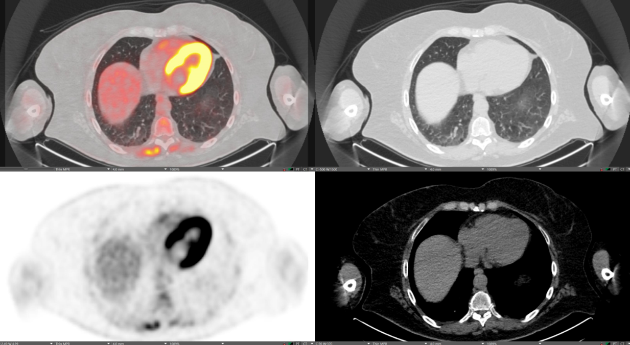
Normal myocardial uptake. Top left: Fused image overlappting PET and CT. Top right: CT in lung window. Bottom left: PET image. Bottom right: CT in soft tissue window.
CT portion can be fused with the PET image to provide high quality anatomic detail for direct comparison of anatomy and physiology.
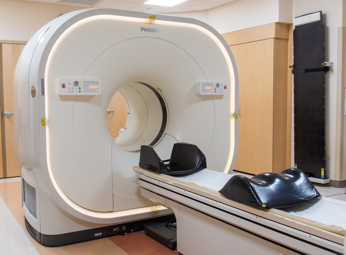
Scanner looks like a normal CT scanner with a built in gamma detector.
PET and CT images can be combined to create a "fused" image (see top left image).
What is considered "normal" depends on which radiotracer is used.
FDG simulates glucose metabolism- organs that highly metabolize glucose will have a higher signal. For example: heart, brain, bone marrow, kidneys…
On the right, a "normal" FDG PET-CT shows bright uptake in the highly metabolic myocardium.
Abnormally high signal where it shouldn't be is a red flag!
For example- pulmonary nodules are very common on CT and it can be difficult to know how worrisome it is by CT alone. On PET-CT, benign pulmonary nodules have little to no uptake, whereas lung cancer will be very bright.