Nuclear Medicine Scanners
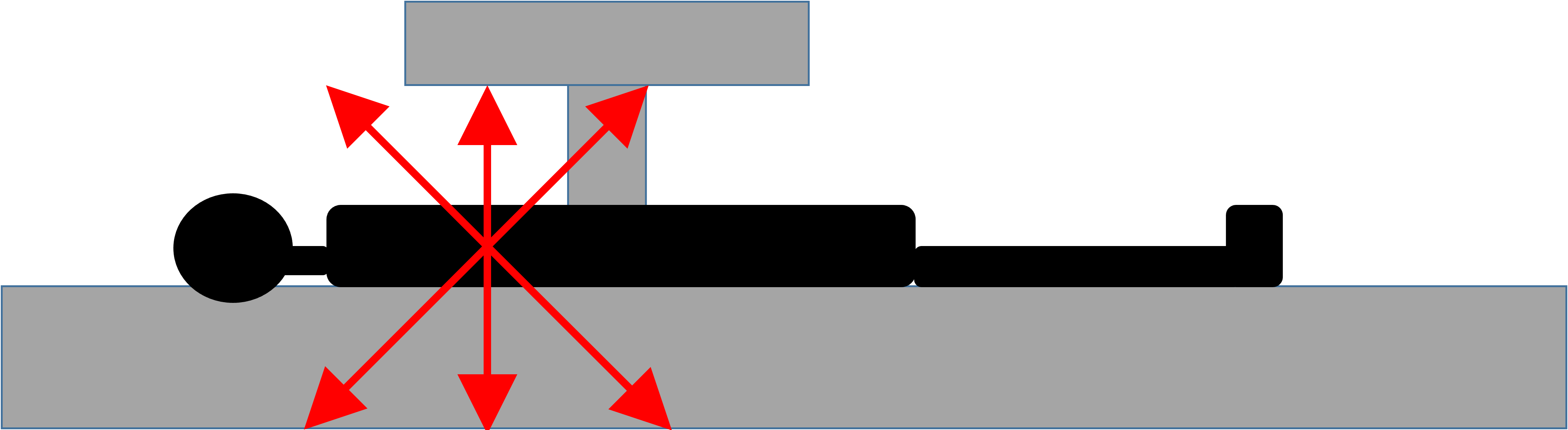
- Nuclear medicine scanners are only detectors that pick up energy emitted from radioisotopes given to patients either orally or intravenously.
- No radiation is emitted from the scanner.
- Scanners count the number of photons and convert the emissions into images.
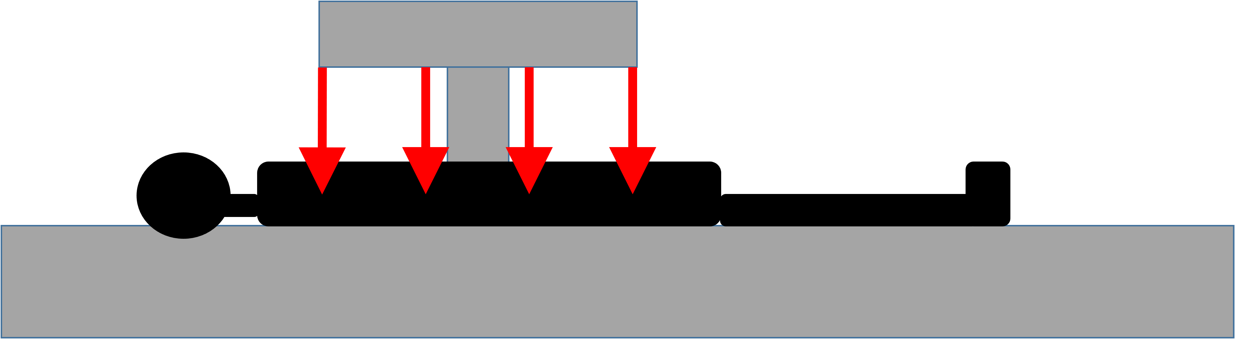
- In x-ray or CT imaging, radiation is externally generated, passes through the patient, and is captured by the detector on the other side.
- In nuclear imaging, the radioactive isotopes are ingested or injected, and subsequent radiation is internally generated and externally detected as radioactive decay occurs.
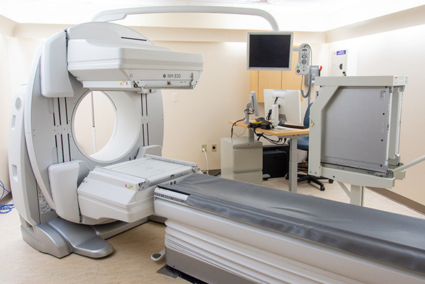
Example of gamma camera
- Positron Emission Topography (PET) scanners are different from gamma cameras.
- They detect positron emitting isotopes.
- Positron = beta particle with positive charge.
- Positron is emitted from the nucleus and after a short distance will collide with an electron, causing an "annihilation" event.
- Annihilation creates two photons traveling in opposite directions which the PET scanner detects and converts into an image.
- PET detector has to detect both photons traveling in opposite directions at nearly the same time for it to be counted.
- PET scanners also include a CT scanner, which can be used to create "fused" images.
Annihilation Event
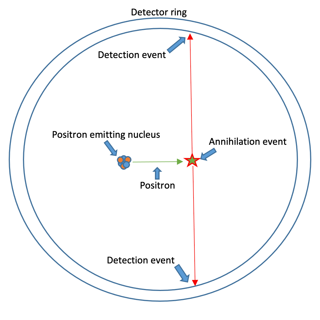
- Positron is emitted from the nucleus.
- Electron and Positron collide, and an annihilation event occurs.
- Two identical photons are generated, traveling in opposite directions.
- Two signals are captured by the detector ring simultaneously.