![]()
Chromatin is double stranded DNA wound
around histone octamers to form nucleosomes
held together by linker DNA. During
Interphase
(G1, S, G2) chromatin is not condensed.
As the cell goes into mitosis or meiosis however, the nucleosomes
are packed together to form chromosomes.
Chromosomes
are first visible in Prophase and become increasingly condensed until metaphase.
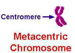
Fully condensed metaphase chromosomes have a centromere
which
divides the chromosome into a p arm (short)
and a q arm (long).
QUESTION: What is the
difference between chromatin and a chromosome?
Homologous chromosomes are usually seen as dyads with two identical chromatids. This is the result of DNA replication and chromosome duplication during S phase of the cell cycle.
QUESTION: What is the
difference homology and identity?
 Each
gene has a locus, or location, on a DNA molecule.
Therefore, when the DNA molecule is packaged into nucleosomes, which
are packaged into chromosomes - each gene has a locus on the chromosome.
The loci of the genes on a chromosome can be indicated on an idiogram
of a chromosome (click
on the image for an enlargement).
Each
gene has a locus, or location, on a DNA molecule.
Therefore, when the DNA molecule is packaged into nucleosomes, which
are packaged into chromosomes - each gene has a locus on the chromosome.
The loci of the genes on a chromosome can be indicated on an idiogram
of a chromosome (click
on the image for an enlargement).
 A
technique called FISH (fluorescent in situ hybridization) allows
a single gene to be visualized (click
on the image for an enlargement).. A copy of
the gene is made, and then labeled with fluorescent dye. Since the
DNA in the copy is complementary to the DNA of the same gene in a chromosome,
it will hybridize to the chromosome at the locus of the gene.
When this is viewed under a microscope, using uv light instead of visible
light, the dye fluoresces yellow and shows the locus of the gene.
In this image the human gene encoding the enzyme muscle glycogen phosphorylase
is seen as a yellow fluorescent spot close to the centromere of each sister
chromatid of two homologous dyads of chromosome 11.
A
technique called FISH (fluorescent in situ hybridization) allows
a single gene to be visualized (click
on the image for an enlargement).. A copy of
the gene is made, and then labeled with fluorescent dye. Since the
DNA in the copy is complementary to the DNA of the same gene in a chromosome,
it will hybridize to the chromosome at the locus of the gene.
When this is viewed under a microscope, using uv light instead of visible
light, the dye fluoresces yellow and shows the locus of the gene.
In this image the human gene encoding the enzyme muscle glycogen phosphorylase
is seen as a yellow fluorescent spot close to the centromere of each sister
chromatid of two homologous dyads of chromosome 11.
![]()
 A
karyotype
is a representation of all the chromosomes in a cell. A typical photographic
karyotype is shown here. To begin with, actively dividing mitotic
cells are treated with colchicine or colcemid.
These drugs inhibit the polymerization of spindle fibers, so the chromatids
cannot separate as they normally would in Anaphase. Instead cells,
which are arrested in metaphase, accumulate. These cells can be swollen
by osmosis in a hypotonic solution.
If they are then dropped from a height onto a glass slide, many of the
cells and nuclei burst, and the chromosomes
splash apart. A "chromosome spread"
from a single nucleus is shown on the right of the image.
These spreads are then stained, usually with Giemsa stain (see below) or
with a fluorescent dye and viewed under the microscope. Photomicrographs
are taken. Each chromosome is then cut out
of the photo, matched with its homologous partner, and pasted onto a piece
of paper to produce the karyotype on the left of the image
above.
A
karyotype
is a representation of all the chromosomes in a cell. A typical photographic
karyotype is shown here. To begin with, actively dividing mitotic
cells are treated with colchicine or colcemid.
These drugs inhibit the polymerization of spindle fibers, so the chromatids
cannot separate as they normally would in Anaphase. Instead cells,
which are arrested in metaphase, accumulate. These cells can be swollen
by osmosis in a hypotonic solution.
If they are then dropped from a height onto a glass slide, many of the
cells and nuclei burst, and the chromosomes
splash apart. A "chromosome spread"
from a single nucleus is shown on the right of the image.
These spreads are then stained, usually with Giemsa stain (see below) or
with a fluorescent dye and viewed under the microscope. Photomicrographs
are taken. Each chromosome is then cut out
of the photo, matched with its homologous partner, and pasted onto a piece
of paper to produce the karyotype on the left of the image
above.
This is a karyotype of a child with Down's Syndrome. What is unusual about this karyotype?
![]()
As you know most organisms are diploid - that is they have
2 sets of chromosomes. BUT how can we tell the homologous
pairs of chromosomes apart? As shown in the figure, there
are 3 ways:
 |
CLICK
on the image for an enlargement!
Size: some chromosomes are large; some small; some
in between.
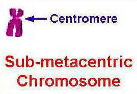
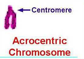
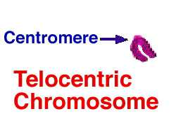
Centromere position: some centromeres are in the middle (metacentrics), and divide the chromosome into 2 equal arms; some centromeres are offset to one end ( sub-metacentrics and acrocentrics) so there is a p arm and a q arm. Some centromeres are placed at the end of the chromosome (telocentrics). |
|
|
Banding pattern: mammalian chromosomes do not naturally exhibit bands. However if they are stained with Giemsa stain, using a special techniques called G-banding, bands can be made to appear. The value of banding is plain by looking at the smaller sub-metacentrics (6 - 12). In the karyotype above the size and position of the centromeres is very similar. However if you look carefully, each of these can be readily distinguished by a different pattern of G bands. The dark bands are thought to be heterochromatin and the lighter bands are thought to be euchromatin.
|
|
 |
CLICK on the image for an enlargement! Each of the bands on a chromosome is distinguished by a numerical code.
This is a cytogenetic
map.
The gene which is defective in Cystic Fibrosis is located at 7q31.2.
This
does not mean the locus of the CF gene is in the thirty-first band!
It means the locus is in the second subdivision
of the first subdivision of region three of the
long arm
of chromosome 7.
|
![]()
The X chromosome carries hundreds of genes but few, if any, of these
have anything to do directly with sex. However, the inheritance of these
genes follows special rules. These arise because:

An idiogram of the human X chromosome showing the G bands. Also shown are the locations of some 'markers' or 'signposts' which are used for mapping. CLICK on the image for an enlargement! |

An idiogram of the human X chromosome showing the G bands. Also
shown are the locations of several genes in which mutant alleles result
in disease phenotypes! Note the difference
in size between the X and Y chromosome.
CLICK on the image for an enlargement! |
Y chromosome morphology
Back to Top of Page. RETURN TO REVIEW
|