![]()
![]()
1. The CHIME Site at the University of Massachusetts
2. Section #1: Hemoglobin and
Heme
a. Viewing the molecule3. Section #2: Hemoglobin Secondary Structurei. Tranlateb. Using the menu to display the molecule
ii. Rotate
iii. Roll
iv. Zoomi. Rotationc. The other buttons in this section of the tutorial
ii. Display
iii. Color
iv. Select
a. Alpha Helix
b. A rainbow coloring scheme from the N-terminus to the C-terminus helps to discern the separate alpha helices.
c. This is a cartoon representation.
d. We'll focus on a single alpha helix. This helix is at the protein-water interface.
e. Here is the isolated alpha helix.
f. The backbone representation connects alpha carbon positions in this alpha helix. These lines do not represent the positions of any actual chemical bonds.
g. Here are the actual bonds of the alpha helix backbone: three atom repeats of nitrogen, alpha carbon, carboxy carbon.
| These resources work best
with Netscape Navigator 4.08-4.76. Netscape 6 doesn't work with Chime 2.
Internet Explorer will give molecular images with Chime in some of the
UMass Resources, but rarely works properly. Use
Netscape!
Chime is a plugin; Netscape invented plug-ins and defined the plug-in interface. The incompatibility of Chime 2 with Internet Explorer is the result of Microsoft never having provided full plug-in support in IE. |
![]()
Go to the CHIME site at the University of Massachusetts.
![]()
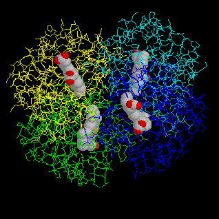
The rotating model of hemoglobin shows several things:
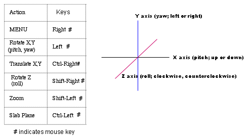 Translate
X or Y (moved left or right; up or down). with the mouse
by holding down the control key and the right button on the
mouse.
Translate
X or Y (moved left or right; up or down). with the mouse
by holding down the control key and the right button on the
mouse.The Select function allows us to display different parts of the molecule in different ways.
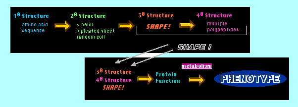
We know that proteins have shape because it is necessary for them to bind something in order to perform their function. For example and enzyme has an active site into which the substrate fits. More generally we refer to these as the binding site on the protein and the ligand which fits into it.
In Hemoglobin we can display the protein chains and the ligand (the Heme group) in different ways. We can display hemoglobin to emphasize the shape of the binding site:
The other buttons in this section of
the tutorial:
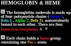
![]() Each chain holds
a heme group containing one Fe++ atom.
Each chain holds
a heme group containing one Fe++ atom.
![]() The heme-iron complexes
are colored red because they give hemoglobin its red color. Actually,
it is the yellow colored atom in the center which gives the Heme group
the red color. This atom is Iron. Heme has a red color because
oxidized iron (rust) is red! This is also why oxygenated blood
is red.
The heme-iron complexes
are colored red because they give hemoglobin its red color. Actually,
it is the yellow colored atom in the center which gives the Heme group
the red color. This atom is Iron. Heme has a red color because
oxidized iron (rust) is red! This is also why oxygenated blood
is red.
![]() Now the heme molecules
have been colored by element. The Heme groups displayed using
CPK colors.
Now the heme molecules
have been colored by element. The Heme groups displayed using
CPK colors.
![]() Spacefill view of
atoms that make up a single heme molecule. The Heme group displayed
to show the true size of each atom, and the real shape of the molecule.
Spacefill view of
atoms that make up a single heme molecule. The Heme group displayed
to show the true size of each atom, and the real shape of the molecule.
![]() Here is how iron
is attached to the rest of the heme molecule.
Here is how iron
is attached to the rest of the heme molecule.
![]() An elemental oxygen
molecule binds to the ferrous iron atom in the lungs where oxygen is abundant,
and is released later in tissues which need oxygen. Note that there
is a difference between a single oxygen atom and an Oxygen molecule
which is composed of two oxygen atoms!
An elemental oxygen
molecule binds to the ferrous iron atom in the lungs where oxygen is abundant,
and is released later in tissues which need oxygen. Note that there
is a difference between a single oxygen atom and an Oxygen molecule
which is composed of two oxygen atoms!
![]() The position of bound
elemental oxygen in one chain of hemoglobin.
The position of bound
elemental oxygen in one chain of hemoglobin.
![]() Space occupied by
the heme bound oxygen in the polypeptide chain.
Space occupied by
the heme bound oxygen in the polypeptide chain.
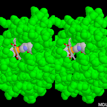
![]() A histidine nitrogen
binds to the iron, helping to anchor its position. Don't worry about
this one!
A histidine nitrogen
binds to the iron, helping to anchor its position. Don't worry about
this one!
![]() A spacefill view
(with the exception of the heme molecule) of the hemoglobin polypeptide
chain. This view shows a stick model of the Heme group with the
yellow colored iron atom in the center. The molecules on either side
displayed in spacefill mode and CPK color are the side chains of 2 amino
acids in the protein chain.
A spacefill view
(with the exception of the heme molecule) of the hemoglobin polypeptide
chain. This view shows a stick model of the Heme group with the
yellow colored iron atom in the center. The molecules on either side
displayed in spacefill mode and CPK color are the side chains of 2 amino
acids in the protein chain.
![]()
Click on the link for "Hemoglobin Secondary Structure".
![]() Most of the amino acids
in hemoglobin form alpha helices.
Most of the amino acids
in hemoglobin form alpha helices.
When you click on the "X" button, the rotating model of the hemoglobin
molecule will load again. Stop the rotation so you can view the
molecule easily. What you see is one of the four polypeptides
and a single heme group (the author does not specify whether this
is an a or b globin
chain).
| REMEMBER from the tutorial above that: "The hemoglobin molecule is made up of four polypeptide chains (Alpha 1, Beta 1 , Alpha 2, Beta 2), non-covalently bound to each other. There are four heme-iron complexes." |
The segments of the amino acid chain which fold into a
helix
are shown in red. Random coil (the segments which do not fold into
a
helix)
are shown in white. Start the rotation again so you can
see the overall 3D structure.
![]() A rainbow coloring
scheme from the N-terminus to the C-terminus helps to discern the separate
alpha helices.
A rainbow coloring
scheme from the N-terminus to the C-terminus helps to discern the separate
alpha helices.
This coloring scheme helps to trace out the chain from the beginning which is colored blue -- light blue -- teal -- green -- yellow -- orange -- red, which is the end.
![]() Here is the isolated
alpha helix.
Here is the isolated
alpha helix.
![]() The backbone representation
connects alpha carbon positions in this alpha helix. These lines do not
represent the positions of any actual chemical bonds.
The backbone representation
connects alpha carbon positions in this alpha helix. These lines do not
represent the positions of any actual chemical bonds.
![]() Here are the actual
bonds of the alpha helix backbone: three atom repeats of nitrogen, alpha
carbon, carboxy carbon.
Here are the actual
bonds of the alpha helix backbone: three atom repeats of nitrogen, alpha
carbon, carboxy carbon.
Stop the rotation. Use the mouse to manipulate the molecule into a vertical position. Reposition the model to the center of your window. Resize the model so that the entire length fits in your window. Using the mouse menu go to Color; then Amino Acid. Now you can see the individual amino acids in the chain which is coiled into the a helix.
![]() Hydrogen bonds (white)
stabilize the alpha helix. Don't worry about this one.
View it if you are interested.
Hydrogen bonds (white)
stabilize the alpha helix. Don't worry about this one.
View it if you are interested.
![]() The sidechains on
the alpha carbons are shown. Don't worry about this one.
View it if you are interested.
The sidechains on
the alpha carbons are shown. Don't worry about this one.
View it if you are interested.
![]() Now the sidechain
elements are identified: C H O N Don't worry about this
one. View it if you are interested.
Now the sidechain
elements are identified: C H O N Don't worry about this
one. View it if you are interested.
![]()
Summary
![]()
Review
![]()
| RETURN to the Review Page |
Go to the top of the page.