|
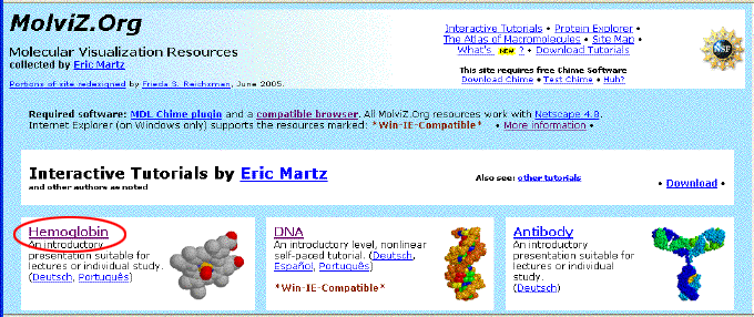
To see what CHIME can do, first
open
a new window. In the new window, go to: "
MolViZ.Org. Molecular Visualization
Resources" (hosted on the San Diego Supercomputer)
.
Then click on
the link to "
Hemoglobin".
The index page
for the Hemoglobin tutorial should appear. Click on the Hemoglobin
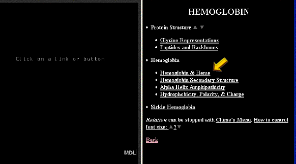
link in large bold text. Now you should see a black
page with 2 frames. The right frame contains links to 3D visualizations
of the various aspects of hemoglobin structure which will be explored.
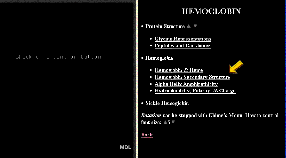
In the right frame of the Hemoglobin Index page, click on "Hemoglobin
& Heme".

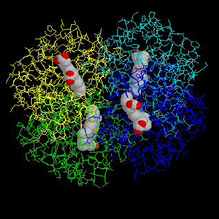
Hemoglobin and Heme

Click on
image to see full-size! |
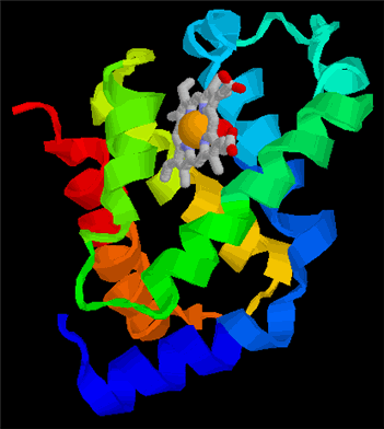
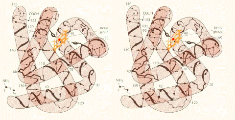
 The
hemoglobin molecule is four polypeptide chains .... The
hemoglobin molecule is four polypeptide chains ....
Click on the "X" button opposite
"The hemoglobin molecule is made up of four polypeptide chains ....".
This should load a rotating model of the hemoglobin molecule in the left
frame. The rotating model of hemoglobin shows several things:
-
It is composed of four separate chains of amino acids (polypeptides).
Each polypeptide is displayed in a different color.
-
Each polypeptide binds one heme group which is displayed in red.
|
|
Click on
image to see full-size! |
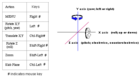
Viewing
the molecule:
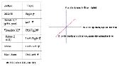
Engineers refer to the position of
an object in 3 dimensions using the terms pitch (movement around the Z
axis), yaw (movement around the Y axis) and roll (movement around the X
axis), as shown in the thumbnail to the left.
The viewer displays different aspects
of the hemoglobin molecule as follows:
-
Translate X or Y (moved left or right; up or down).
with the mouse by holding down the control key and the right
button on the mouse.
-
Yaw and Roll. The image can be
yawed (rotated left and right about the Y axis ) or rolled (rotated up
or down about the X axis) with the mouse by holding down the left
button on the mouse.
-
Pitch. The image can be pitched
(rotated clockwise or counterclockwise) about the Z axis with the mouse
by holding down the shift key and the right button of the
mouse.
Zoom. The image can be resized
with the mouse by holding down the shift key and the left button
on the mouse.
|
|
Click on
image to see full-size! |
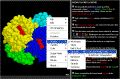
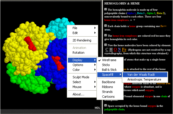
Display
Menu.
A menu for changing the display is
accessed by holding down the right button of the mouse
-
Rotation: This function can be
toggled on or off.
-
Display: The "Display"
menu allows us to view different aspects of the structure:
-
Hold down the right mouse button. Choose "Display" --- "Spacefill"
--- "Van der Waals Radii".
-
Resize the molecule by holding down the shift key and the left mouse button.
Move the mouse up or down to zoom in or out. Reduce the molecule
to the height of your window.
-
Manipulate the molecule with your mouse to stand it up vertically.
-
Hold down the right mouse button. Choose "Options" --- "Stereo Display".
-
Reset:
Click on the "X" button opposite "The hemoglobin molecule is made up of
four polypeptide chains ...."
|
|
Click on
image to see full-size! |
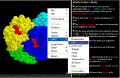
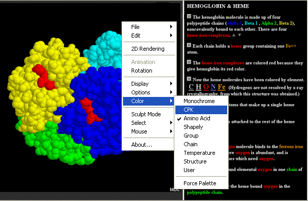
Color
Menu.
The following options are available for color display. Play with
them.
-
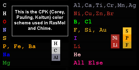
Monochrome - self explanatory
-
CPK (carbon grey; hydrogen white; oxygen red; nitrogen blue; phosphorus
gold)
-
Amino Acid - every amino acid is displayed in a different color
-
Shapely - the amino acids are displayed according to the chemical
properties of their side chains
-
Group - this emphasizes the different elements of secondary structure
and the ends of each polypeptide chain. It is most useful when used
in the "ribbon" or "cartoon" display mode.
-
Chain - each of the polypeptide chains is displayed in its own color
-
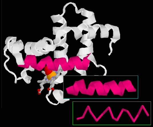
Structure - this emphasizes the different secondary structures.
Alpha helix is displayed in rose; beta pleated sheet in gold; random coil
in thin blue/white lines. It is most useful when used in the "ribbon"
or "cartoon" display mode.
Temperature and User: not useful for our purposes.
|
|

Click on
image to see full-size! |
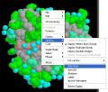
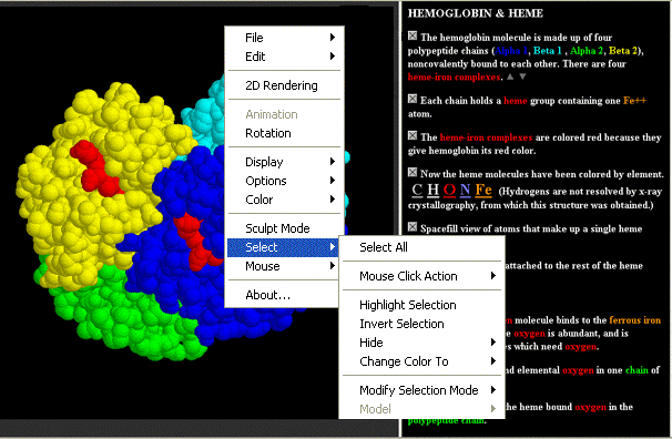
The
Select
Menu displays different parts of the molecule in different ways.
We know that proteins have shape because it is necessary for
them to bind something in order to perform their function.
For example and enzyme has an active site into which the substrate
fits. More generally we refer to these as the binding
site on the protein and the ligand
which fits into it.
In Hemoglobin we can display the protein chains and the ligand (the
Heme group) in different ways. We can display hemoglobin to
emphasize the shape of the binding site:
-
Hold the mouse button down. Go to Select; then go to Hetero;
then go to Ligand ("hetero" refers to any other atoms and
molecules which are not part of the polypeptide chains).
-
Go to the Color menu and select "CPK"
-
Go back to Select; then go to Protein.
-
go to the Display menu and select "Wireframe"
-
You should now see something like this.
Move one of the Heme groups to the center of the screen. Zoom
in; note how the atoms in the side chains of the amino acids closely
contact the Heme group. They form a binding site which holds the
Heme group in. Now
you can see why SHAPE is so important!!!! |
|
| The other buttons in this section
of the tutorial are described below. Feel free to take a quick look at these:
Each chain holds
a heme group containing one Fe++ atom.
Heme group = porphyrin
ring (colored in red) + Fe2+
atom chelated in the center.
There are 4 hemes because there are 4 chains.
The heme-iron complexes
are colored red because they give hemoglobin its red color. Actually,
it is the yellow colored atom in the center which gives the Heme group
the red color. This atom is Iron. Heme has a red color because
oxidized iron (rust) is red! This is also why oxygenated blood
is red.
Now the heme molecules
have been colored by element. The Heme groups displayed using
CPK
colors.
Spacefill view of
atoms that make up a single heme molecule. The Heme group displayed
to show the true size of each atom, and the real shape of the molecule.
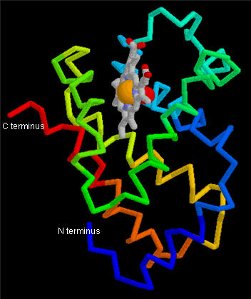
Here is how iron
is attached to the rest of the heme molecule. Notice that the Fe2+
is coordinately bound by 4 heterocyclic ring nitrogens.
An elemental oxygen
molecule binds to the ferrous iron atom in the lungs where oxygen is abundant,
and is released later in tissues which need oxygen. Note that there
is a difference between a single oxygen atom and an Oxygen molecule
which is composed of two oxygen atoms!
The position of bound
elemental oxygen in one chain of hemoglobin.
Space occupied by
the heme bound oxygen in the polypeptide chain. Notice how close is
the complementary fit between the heme group and the cavity formed by the
alpha helices in the beta globin chain.
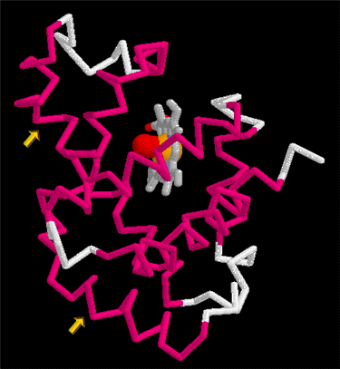
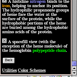
A histidine nitrogen
binds to the iron, helping to anchor its position. Actually,
there are 2 histidine residues shown here!
-
Histidine 63 (on the right side) binds
to the O2 molecule, stabilizing it.
-
Histidine 92 (on the left side) binds to the
Fe2+ atom, holding it in place from the other side.
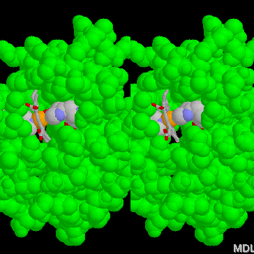
A spacefill view
(with the exception of the heme molecule) of the hemoglobin polypeptide
chain. This view shows a stick model of the Heme group with the
yellow colored iron atom in the center. The molecules on either side
displayed in spacefill mode and CPK color are the side chains of 2 amino
acids in the protein chain.
-
Hold down the button on your mouse and go to the Options menu.
At the bottom choose Stereo Display. Adjust the size to look
something like this.
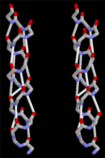
Put your nose close up to
the screen, and move slowly backward concentrating on the "third" image
in the center. At some point the image should appear 3 dimensional!
This view gives you a good idea of how closely ligands fit into binding
pockets .......... and how important SHAPE is to FUNCTION!!!
|
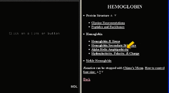
Secondary Structure
At the bottom of the HEMOGLOBIN & HEME tutorial is a "Back"
button. This will return you to the first
page. Click on the link for "Hemoglobin
Secondary Structure".
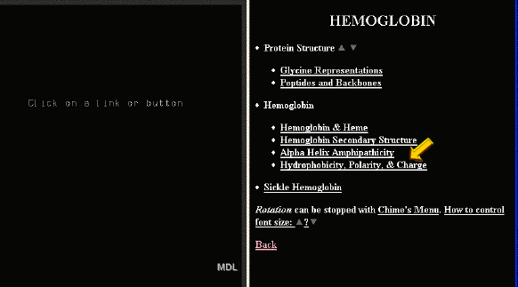
Amphipathicity of the Alpha Helix
At the bottom of the Hemoglobin Secondary Structure tutorial is
a "Back" button. This will return
you to the first page. Click on the
link for "Amphipathicity of the Alpha
Helix".
From your knowledge of Organic Chemistry, what does "amphipathic"
mean?
Hydrophobicity, Polarity and Charge
At the bottom of the tutorial is a "Back" button.
This will return you to the first page.
Click on the link for "Hydrophobicity
polarity and charge".
|
Click on
thumbnail to see full-size! |
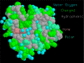
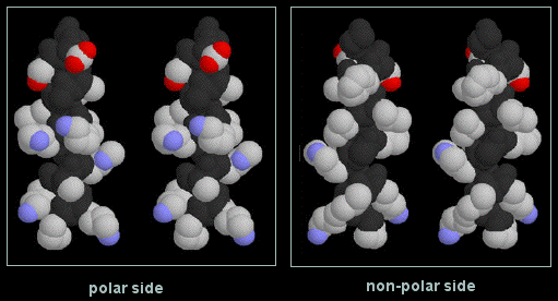
"This view of the beta chain....."
This graphic shows a space-filling
model of a beta globin chain with the polar residues in blue or green,
and the non-polar residues in gray. The heme group is colored light pink.
Turn the stereo option on. Turn on rotation.
When viewed in stereo, do the hydrophilic residues appear more often
on the surface, and the hydrophobic residues more often on the interior? |
|
Click on
thumbnail to see full-size! |
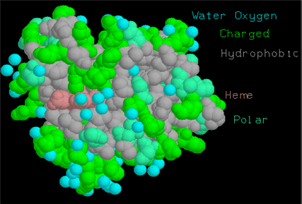
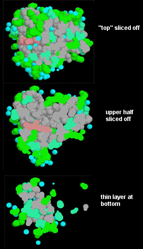
Protein Explorer allows the user
to view slices through the molecule, in much the same way as MRI scans
allow physicians to see slices through a patient's body.
Open the menu. Select "Options", then "Slab Mode", as shown in the
thumbnail to the left.
Now hold down both the Control and Left mouse keys, and move the
cursor up or down to slice through the molecule. The views should look
something like this.
The Slab option clearly demonstrates how hydrophobic residues are shielded
by the hydrophilic residues from the aqueous environment surrounding the
molecule. |
|
 Send
us questions or comments!
Send
us questions or comments!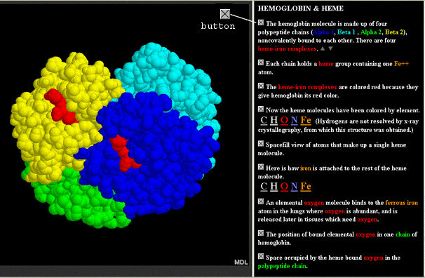
