
CHIME is an application which allows you to view an organic chemical
or biochemical in 3-dimensions, from any angle. It also allows you
to display it in various ways and in different color schemes.
In
order to make the best use of this tutorial, keep two windows open
on your Desktop!
-
In the first window, view this
tutorial. Even though you may have a hard copy, you can't follow the links
unless you have this page open in your browser.
-
In the second window, run Protein
Explorer with the DNA model.
|
To see what CHIME can do, first
open
a new window. In the new window, go to: "
MolViZ.Org. Molecular Visualization
Resources" (hosted on the San Diego Supercomputer)
.
Then click on the
link to " DNA".
The index
page for the DNA tutorial should appear. Scroll down this page until
you see a set of images which looks like this.

The Sugar-Phosphate backbone
Explore the sugar-phosphate backbone by clicking on "C.
Strands and helical backbone" . Now
you should see a model of the double stranded DNA molecule. One strand
is rendered in gold; the other is rendered in brown. Below the model is
a control panel with "X" buttons.
-
Click on the "trace" button to identify
the two sugar-phosphate chains. Click on the "off" button.
-
Click on the "phosphorus" button. This
emphasizes
how the dexoxyribose sugars are linked together
by phosphate molecules.
Click on the "off" button.
-
Click on "X spin" button.
-
Click on the "sugars" button to erase
the sugars, and view the nitrogenous base pairs.
-
A-T base pairs are held together by
two
H-bonds.
-
G-C base pairs are held together by
three
H-bonds.
-
Click on the "Reset" button to restore
the original view. Click on the "trace" button. Click on the "Y spin" button
to rotate the molecule around the Y axis. As you look down through the
molecule from the top, note how the 2 strands twist
around each other to form a hollow tube, with all the base pairs
are to the inside of the helix.
-
Click on the "Reset" button to restore
the original view. Click on the "H bonds" button to erase the H-bond depictions.
erase the dark brown strand with the appropriate button. Hold down the
left button and use the mouse to move the molecule around.
-
Note how
the phosphates connect to the 5' and 3' carbons
in the deoxyribose sugars of each nucleotide.
-
Note how
the nitrogenous bases connect to the 1' carbons in the deoxyribose
sugars of each nucleotide.
The Nucleotides.
Explore the way the nucleotides are linked together by clicking on "D.
Ends, Antiparallelism" . Now
you should see a model of the double stranded DNA molecule with the
sugar phosphate backbone emphasized. Now the molecule is rendered in CPK
color scheme (first introduced by Corey, Pauling
and Koltun)
-
Click on the "5' end" button. Hold down
the left button and drag the mouse to show the 5' end of this DNA in different
orientations.
-
Identify where each nucleotide.
begins and where it ends.
-
Identify the 5' carbons in each of the
first 3 nucleotides.
-
Identify the 3' carbons in each of the
first 3 nucleotides.
-
Identify the 1' carbons in each of the
first 3 nucleotides.
-
What is the name of the base in the
first nucleotide?
-
Why is this called the 5' end of the
strand?

-
Click on the "Reset" button to restore
the original view. Click on the "3' end" button. Hold down the left button
and drag the mouse to show the 3' end of this DNA in different orientations.
-
Identify where each nucleotide.
begins and where it ends.
-
Identify the 5' carbons in each of the
last 3 nucleotides.
-
Identify the 3' carbons in each of the
last 3 nucleotides.
-
Identify the 1' carbons in each of the
last 3 nucleotides.
-
What is the name of the base in the
last nucleotide?
-
Why is this called the 3' end of the
strand?
The Nitrogenous Base Pairs.
Explore the nitrogenous base pairs by clicking on "B.
The Code". Now
you should see a model of the double stranded DNA molecule. Each
sugar-phosphate backbone is colored brown. The nitrogenous bases are colored
red, green, yellow and blue Below the model is a control panel with
"X" buttons.
-
Click on the "1/2" button. Click on the "Spacefill" button. Then click
on the "X spin" button to rotate the molecule around the X axis. Locate
the major groove and the minor
groove. Click on the "Reset" button to restore the original view.
-
DNA contains 4 nitrogenous bases: Adenine;
Guanine; Thymine; Cytosine. Why are they
called nitrogenous bases?
-
Click on the "AT" button. Adenine pairs with Thymine
to form A_T base pairs
with 2 H-bonds.
Move
it around so you can get a good idea of what the base pairs look like.
-
How can you tell which one of the base pairs is the pyrimidine?
-
How can you tell which one of the base pairs is the purine?
-
Identify the 1', 3' and 5' carbons on both deoxyribose molecules.
-
Click on the "GC" button. Guanine pairs with Cytosine
to form G-C base pairs
with 3 H-bonds.
-
How can you tell which one of the base pairs is the pyrimidine?
-
How can you tell which one of the base pairs is the purine?
-
Identify the 1', 3' and 5' carbons on both deoxyribose molecules.
-
Click on the "Replication" button to see an animation of DNA Replication.
-
Click on the "Codons" button to see an animated explanation of how DNA
sequence codes for protein primary structure.
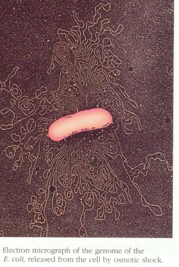
Length of Double Stranded DNA
In living organisms, DNA is a very
long molecule.
-
The bacterium Escherichia coli, which
lives in our guts, has a chromosome 4,000,000 base pairs long (4 megabases).
Here is a scanning electron micrograph of a burst bacterium
with its DNA spilling out !
-
An average human chromosome has 150,000,000
base pairs (and there are 46 chromosomes in each one of our cells!).
Watson and Crick
A double stranded DNA molecule is a helix.
Imagine holding a toy ladder in your right hand. Then pointing
it away from you, twist the top with your
left hand in a
clockwise
manner. This produces what is known as a
right-handed
helix.
The B-form
of DNA is a right handed helix. It is the classical structure first
described by James Watson and Francis Crick.
|