First step is to determine if the study is technically adequate.
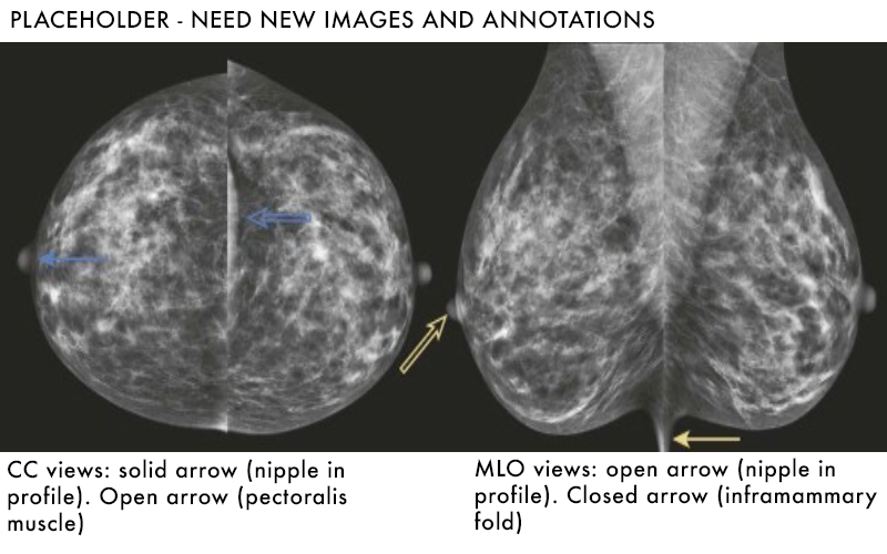
Nipple should be in profile in at least one view.
Image should be free of motion or artifact.
Adequate breast tissue should be included.
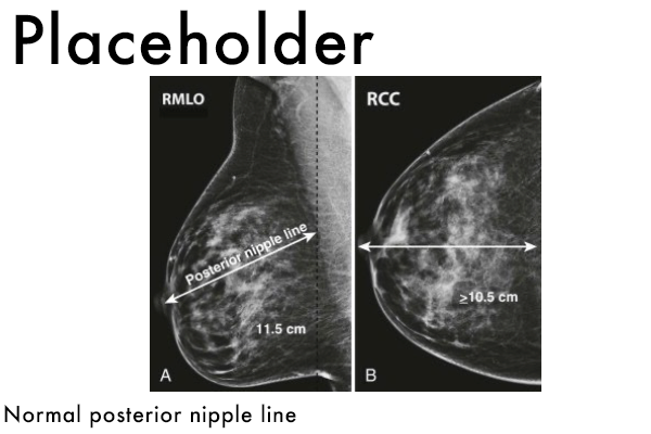
Posterior nipple line: line drawn from posterior nipple on MLO view should intersect with the pectoralis muscle. Line is also measure on CC view, measurements should be within 1.0cm of each other.
Inclusion of the inframammary fold on the MLO view.
Inclusion of retroglandular fat
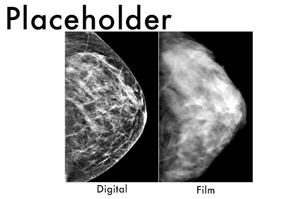
Digital Mammography
Film was used up until 2000, until FDA approved use of digital mammography.
No longer use actual film as the detector, rather a digital detector converts photons into a digital signal.
Digital signal can be viewed on high resolution monitors instead of older light boxes.
Digital mammography has nearly completely replaced film at this point.
Digital mammography also allows use of tomosynthesis (3-D mammography).
Thought to help aid in detection of subtle lesions.