How Images Are Obtained
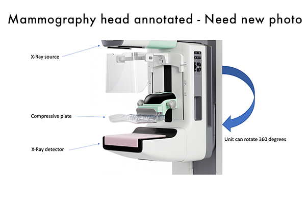
- Breast is compressed between a plastic compression plate and the X-ray detector.
- Why compression is used:
- Compression helps spread out the fibroglandular tissue making it easier for radiologists to detect abnormalities.
- Limits motion artifact which also helps aid in detection.
- More compression means less radiation exposure required to produce optimal images.
- Unit can rotate 360 degrees to obtain different views.
- Cranial-Caudal (CC) and Mediolateral Oblique (MLO) are the standard views obtained on a routine screening mammogram.