![]()
Click on
the image
|
The amino acid sequence (primary structure)
of a globin and b
globin chains. The a
globin sequence is on top; the b
globin sequence is on the bottom (Strickberger, MW.
Genetics, 3rd ed., Macmillan, 1985. p 539). The amino acid
sequences of a globin and
b
globin are similar.
Notice that the 6th amino acid in b globin is glutamic acid!
|
|
|
|
Click on
the image
|
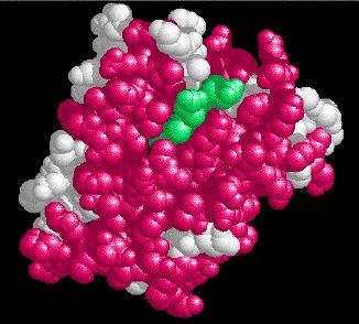
The secondary
and tertiary structure of b
globin is determined by the primary
structure (the amino acid sequence). In the image to the left, the amino
acids have folded into regions of secondary structure - alpha
helix and random coil.
A heme group is composed of a porphyrin ring (depicted in gold) complexed with an iron atom (shown in red). The heme group is held inside a cavity formed by the tertiary structure of the b globin. It is pinned in place by the sidechains of two histidines as shown. When the molecule is depicted in a space-filling model, the 3-dimensional SHAPE can be seen. This is referred to as tertiary structure. In this image, alpha helix is colored magenta, random coil is white, and the heme group is light green. |
|
|
|
Click on
the image
|
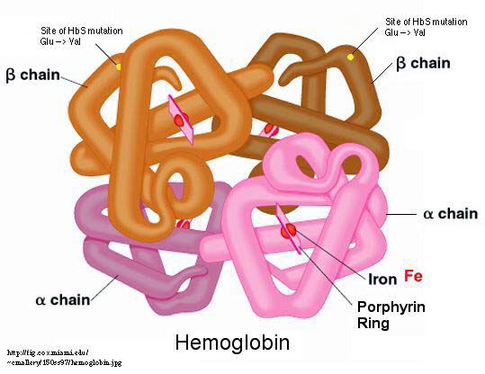
The quaternary structure
of Hemoglobin (HBB) is a tetrameric protein composed of 2 b
globin chains and 2 a
globin chains. b
globin is a peptide chain which contains 147 amino acids. It folds in such
a way as to bind a porphyrin ring with an iron atom (see the link below).
In the graphic to the left, the 2 a globin chains are in black; the 2 b globin chains are in red. The iron atoms are shown as orange spheres, with the porphyrin rings depicted as disks. A space-filling model of hemoglobin, shows the true shape of the protein and the porphyrin rings. The two a globin chains are in different shades of green; the two b globin chains are in different shades of blue. |
|
|
|
Click on
the image
|
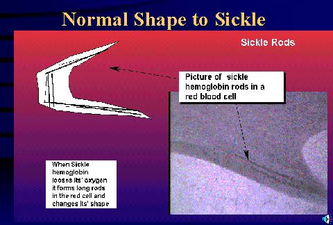
Because valine is substituted for glutamic acid at the 6th position, the b globin chains become "sticky". This causes the hemoglobins to polymerize into long chains. |
|
|
|
Click on
the image
|
The hemoglobin fibers are stiff, and distort the erythrocyte into a sickled shape. In the image to the left, a diagram of a sickled cell is in the upper left, and a micrograph of a sickled cell is in the lower right. Polymerized hemoglobin rods can be seen inside the erythrocyte. |
|
|
|
Click on
the image
|
Many other b globin variants have been found in studies of blood samples from people all over the world. This illustration (from Strickberger, MW. Genetics, 3rd ed., Macmillan, 1985. p 540) shows a human b globin chain. Different amino acids in the chain are labeled with the name of a city. This indicates that a person was found in the city referenced who had an amino acid change at that position in the peptide chain. The wild type amino acid and the amino acid which is substituted in the variant are also indicated. |
|
|
![]()
Back To "Introduction to OMIM"
RETURN to the SITE MAP