Applications
- Evaluation of the gastrointestinal (GI) tract
- Evaluation of the genitourinary tract
- Guidance for arthrograms or joint aspirations
- Guidance for percutaneous interventional procedures - this will be covered in IR lecture
- Guidance for endovascular procedures - arterial, neuro, venous- this will be covered in IR lecture
- Intraoperative guidance- Ortho, GU, GI
- Swallowing mechanism dysfunction
GI Tract
Fluoroscopy is especially useful for the GI system as peristalsis can be observed. It can also demonstrate obstruction and dysfunction of the GI tract in real time, helping to pinpoint the location of the abnormality. Double contrast studies offer similar luminal evaluation as direct visualization (ie: endoscopy or colonoscopy), without the invasive procedure.
Esophagram - for dysphagia, GERD, odynophagia, foreign body.
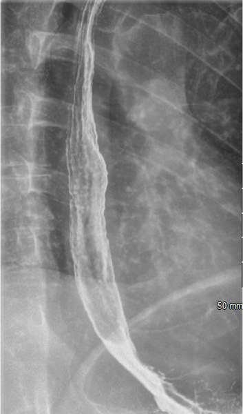
This is an evaluation of the cervical and thoracic esophagus, done with single or double contrast. The above picture is double contrast, done with air and barium.
Upper GI - Pre-op for gastric bypass, regurgitation of food, pain
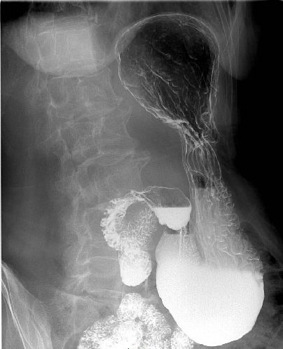
This is an evaluation of the thoracic esophagus, stomach, and duodenum, typically performed with double contrast, by having the patient drink an effervescent solution followed by barium. The above image is a double contrast image of the stomach.
Barium enema - usually for obstruction or to assess surgical anastomosis
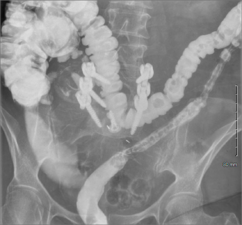
This is an evaluation of the colon, performed by inserting a catheter into the patient's rectum and injecting contrast in a retrograde fashion to the level of the cecum. This can be done with either single or double contrast. The above image was done with single contrast only, water soluble contrast.
Post-operative-GI
Post-gastric sleeve: This single contrast (water soluble) image demonstrates normal flow of contrast through the post-operative stomach, with no leaking beyond the lumen.
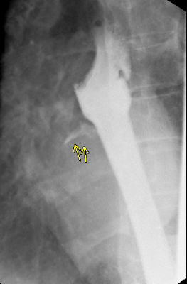
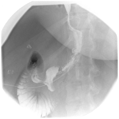
Post-esophageal stent leak: This single contrast (water soluble) image demonstrates a small leak in the esophageal stent (arrow) as shown by contrast going outside of the normal lumen
Small Bowel
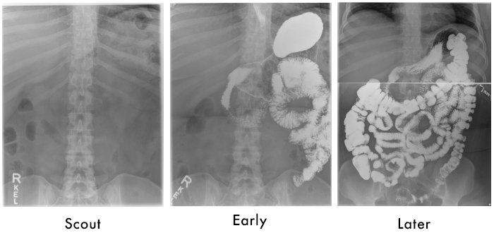
These scout (pre-contrast), early and late post contrast images demonstrate contrast flowing through the stomach and duodenum into the jejunum and ileum. The late image demonstrates contrast throughout the entire colon, proving that there is no small bowel obstruction.
Genitourinary
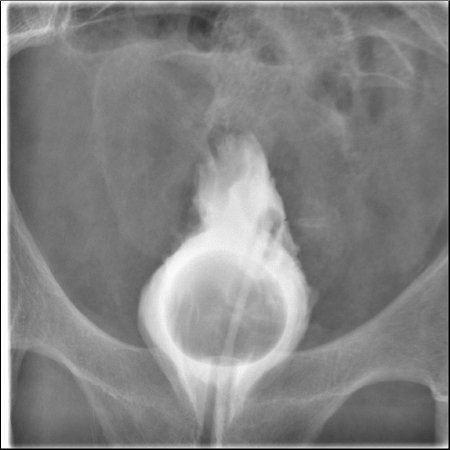
Cystogram- An evaluation of the bladder performed by instilling contrast through a foley catheter. This is typically pre or post op, to assess for ureteral reflux, ureterocele.
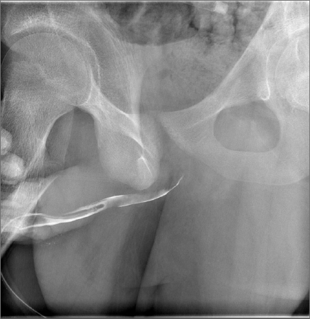
Retrograde urethrogram- An evaluation of the male urethra, performed by instilling contrast through a partially inserted foley catheter, usually after trauma to assess for discontinuity.
Musculoskeletal
Arthrogram- contrast for CT or MRI
- Joint injection w/ corticosteroid- fluoroscopy guidance
- Joint aspiration – to drain effusion/ get sample for micro- fluoroscopy guidance
- A needle is placed into the joint under fluoroscopy, with the use of contrast to make the joint space or bursa visible.

Video - Modified barium for swallowing disfunction
![]()
This is a fluoroscopic video of a pediatric patient being evaluated for swallowing dysfunction by speech and language pathology.