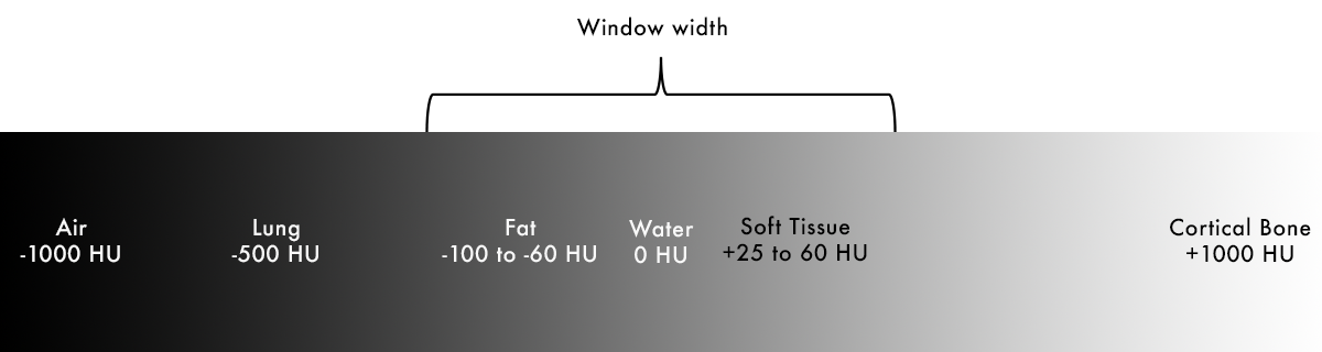
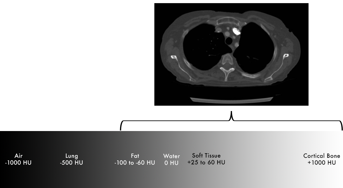
Windowing
- Focuses our gray scale to the tissues of interest – this is the "window width"
- Highlights subtle differences in tissues
- All densities below the specified values are black
- All densities above the specified values are white
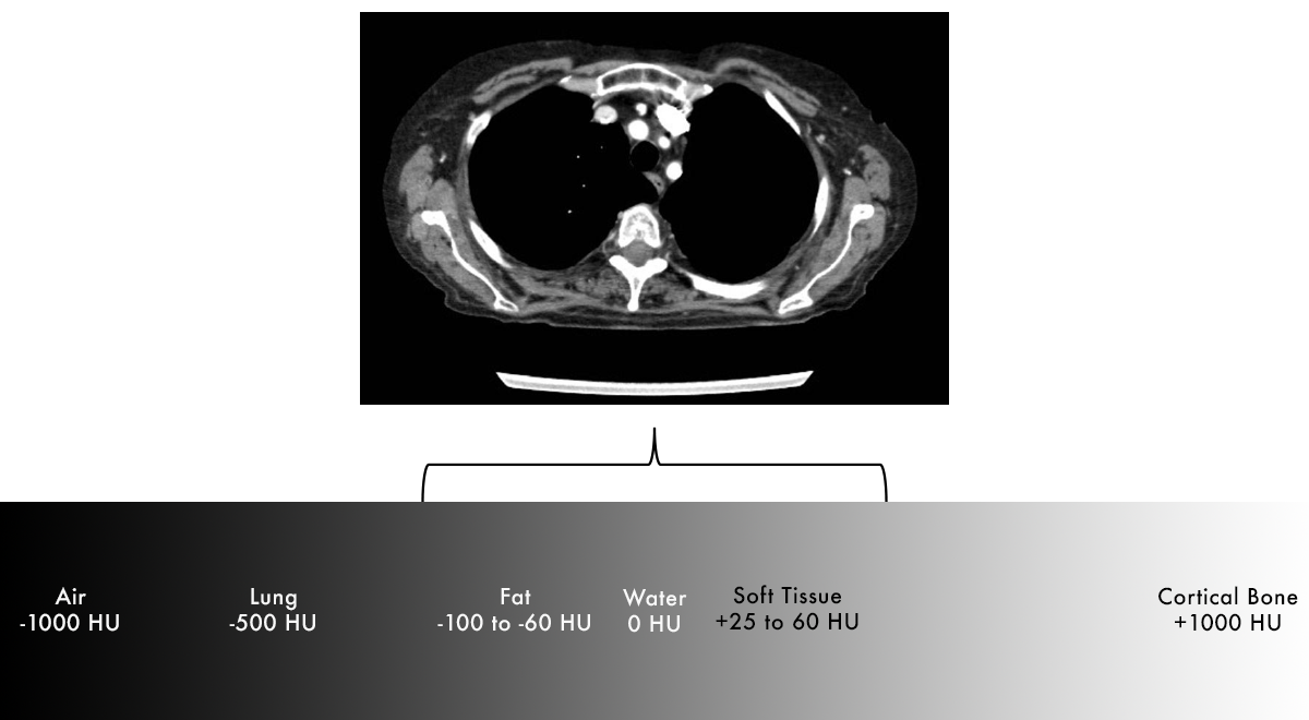
Soft Tissue Window
- It is now very easy to tell the difference between fat (dark grey) and soft tissue (light grey)
- Notice that air (outside of patient) and lungs look exactly the same because they are outside of our specified window of HU values
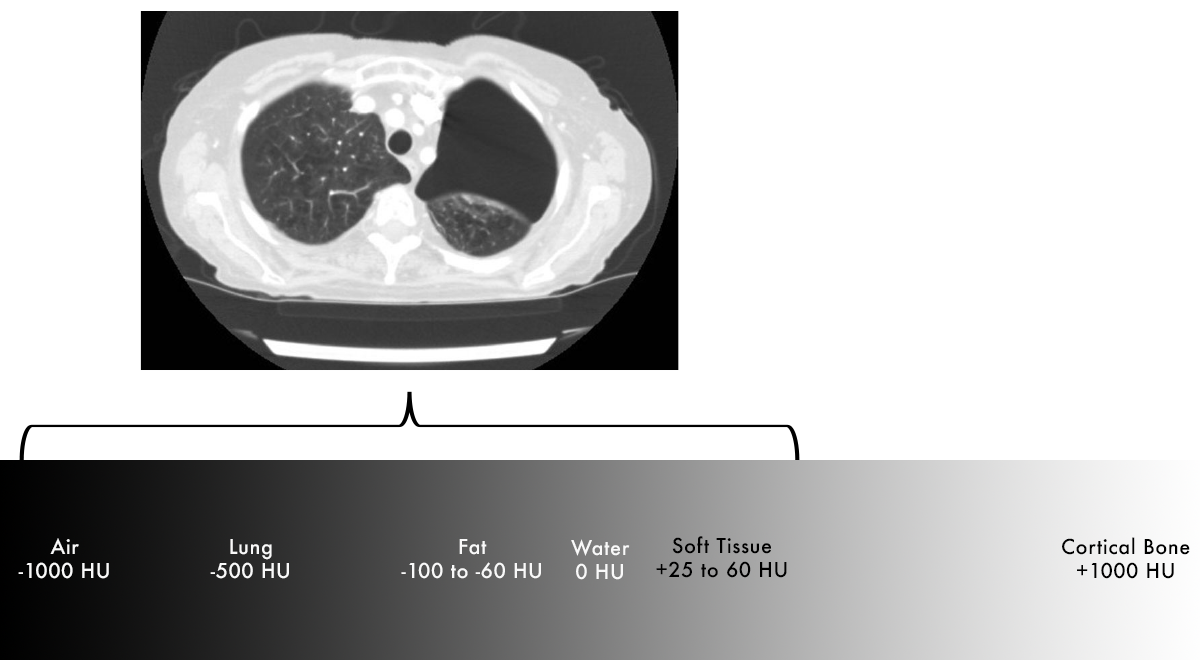
Lung Window
- On the previous image we were unable to see the large pneumothorax
- Can now easily differentiate lung tissue from air in pneumothorax
Bone Window
- Can see differences in bony cortex and medulla
- Easier to detect fractures
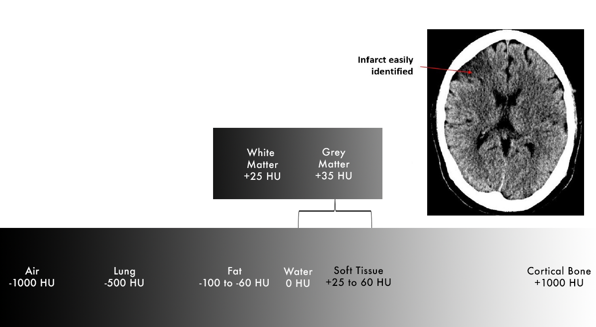
Brain WIndow
- Window can be set narrowly enough that we can distinguish the very small differences in white matter and grey matter
- White matter is slightly less dense than grey matter due to presence of lipid-containing myelin
BRAIN IMAGE NEEDS TO BE REPLACED WITH ONE FROM PACS